==============================================
🎥 Check Out All Videos at Once! 📺
👉 Visit Visualizing MSK Blog to explore a wide range of videos! 🩻
https://visualizingmsk.blogspot.com/?view=magazine
📚 You can also find them on MSK MRI Blog and Naver Blog! 📖
https://www.instagram.com/msk_mri/
Click now to stay updated with the latest content! 🔍✨
==============================================
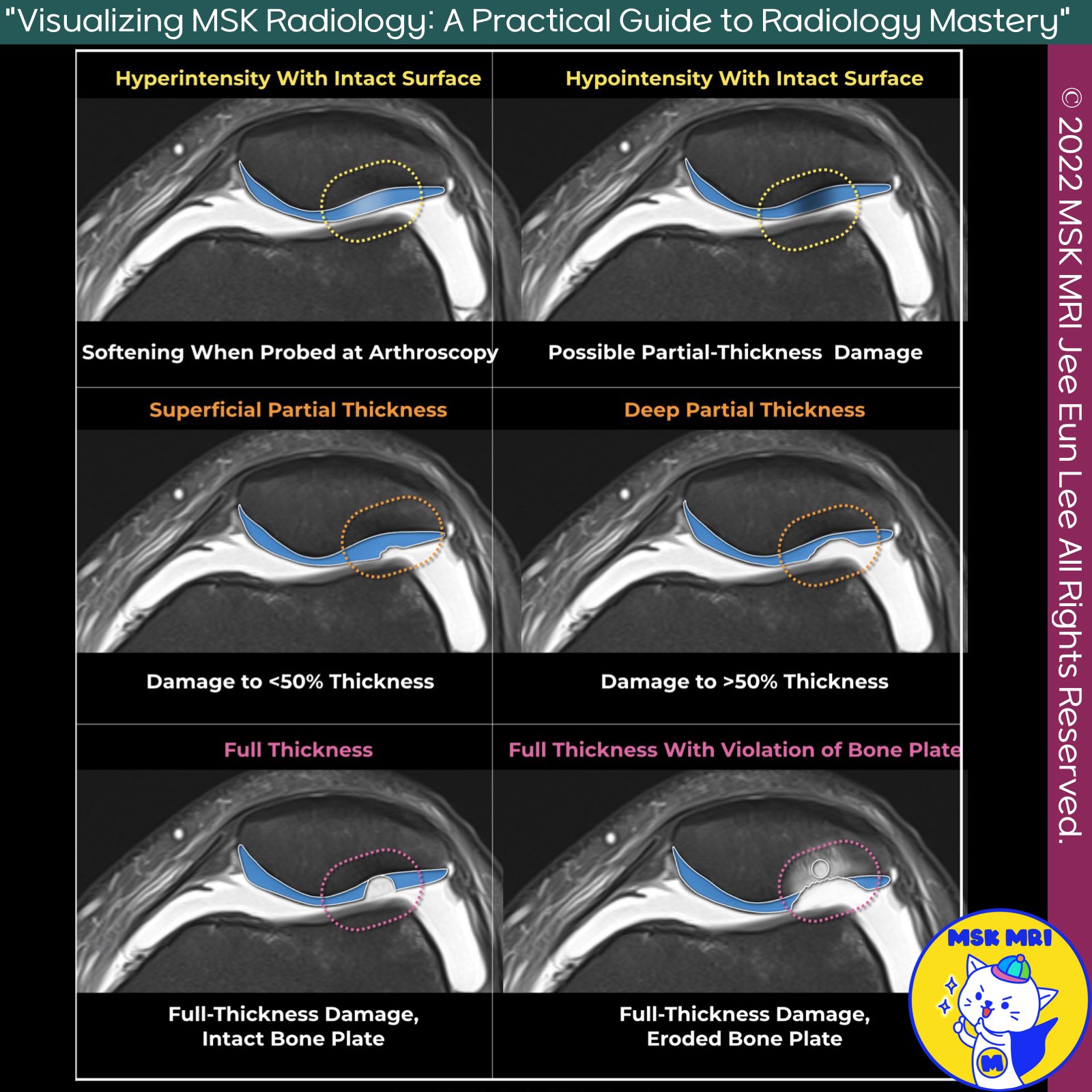
📌MRI Findings in Cartilage Damage
✅ Depth of Cartilage Damage
Modern surgical and MRI grading systems categorize the depth of cartilage damage into four major categories:
- Cartilage Softening: The lowest category.
- Less than 50% Damage: Damage involving less than 50% of the cartilage thickness.
- Greater than 50% Damage: Damage involving greater than 50% of the cartilage thickness.
- Full-Thickness Damage: Complete damage through the cartilage thickness.
✅ Cartilage Defect and Signal Intensity Changes
- In addition to localized loss of cartilage height, signal intensity changes may indicate composition changes or morphologic lesions not visible in standard clinical MRI sequences.
- Both increases and decreases in signal intensity on T2-weighted MR images can be seen with cartilage degeneration at arthroscopy, potentially progressing to morphologic defects on subsequent MRI.
✅ Hyperintense Cartilage Lesions
- Hyperintense lesions in morphologically normal cartilage have a 94% positive predictive value for detecting degeneration at knee arthroscopy.
- These lesions are typically ill-defined, located in abnormally expanded cartilage, and correlate with softening areas at arthroscopy.
- They are due to increased water content, decreased macromolecular content, and disruption of the collagen matrix ultrastructure.
✅ Hypointense Cartilage Lesions
- Hypointense lesions in morphologically normal cartilage have a 64% positive predictive value for detecting degeneration at knee arthroscopy.
- These lesions are often in the middle zone, appearing linear or globular, and are common areas of fibrillation or fissuring at arthroscopy.
- They are due to factors such as disruption of tissue anisotropy, magnetization transfer effects, or mature fibrocartilage.
Reference
RadioGraphics. (2022). Depth of cartilage damage and grading systems. RadioGraphics, 42, 1457–1473
"Visualizing MSK Radiology: A Practical Guide to Radiology Mastery"
© 2022 MSK MRI Jee Eun Lee All Rights Reserved.
No unauthorized reproduction, redistribution, or use for AI training.
#MRI #CartilageDamage #Orthopedics #Radiology #KneeArthroscopy #MedicalImaging #HyperintenseLesion #HypointenseLesion #T2WeightedMRI #CartilageDegeneration


'✅ Knee MRI Mastery > Chap 5AB. Chondral and osteochondral' 카테고리의 다른 글
| (Fig 5-A.11) Hypointense Lesion in Cartilage (0) | 2024.07.05 |
|---|---|
| (Fig 5-A.10) Cartilage Damage Terminology (0) | 2024.07.04 |
| (Fig 5-A.08) International Cartilage Repair Society grade 3 and 4 (0) | 2024.07.04 |
| (Fig 5-A.07) Partial Thickness Cartilage Damage (0) | 2024.07.04 |
| (Fig 5-A.06) Acute Traumatic and Degenerative Cartilage Lesions (0) | 2024.07.04 |