==============================================
🎥 Check Out All Videos at Once! 📺
👉 Visit Visualizing MSK Blog to explore a wide range of videos! 🩻
https://visualizingmsk.blogspot.com/?view=magazine
📚 You can also find them on MSK MRI Blog and Naver Blog! 📖
https://www.instagram.com/msk_mri/
Click now to stay updated with the latest content! 🔍✨
==============================================
📌Chondral Delamination in Cartilage
✅ Definition and Mechanism
- Cartilage delamination separates the articular cartilage at the tidemark from the underlying subchondral bone.
- This occurs due to the discrepancy in compressive stiffness between the noncalcified cartilage and the much stiffer underlying calcified cartilage and bone.
- The lateral deformation of the articular cartilage is constrained by the calcified cartilage bed, leading to shear forces at the tidemark, causing delamination.
- The tidemark delaminates more easily than the deeper junctional region between cartilage and subchondral bone.
- This injury may appear intact at arthroscopy and be seen only as a bulge of the cartilage surface. In contrast to cartilage flaps, where the cartilaginous surface is always violated, delamination injuries may not fit easily into a chondral lesion grading system.
✅ MRI Appearance
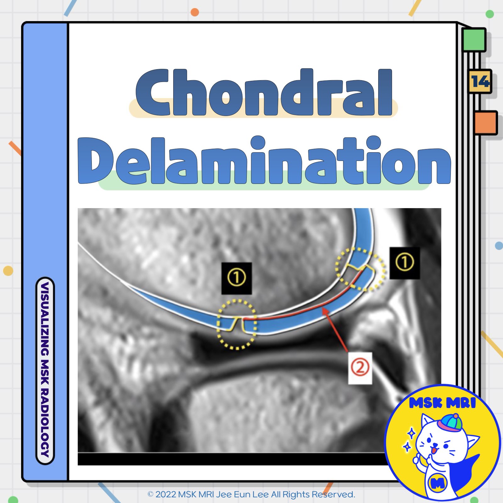
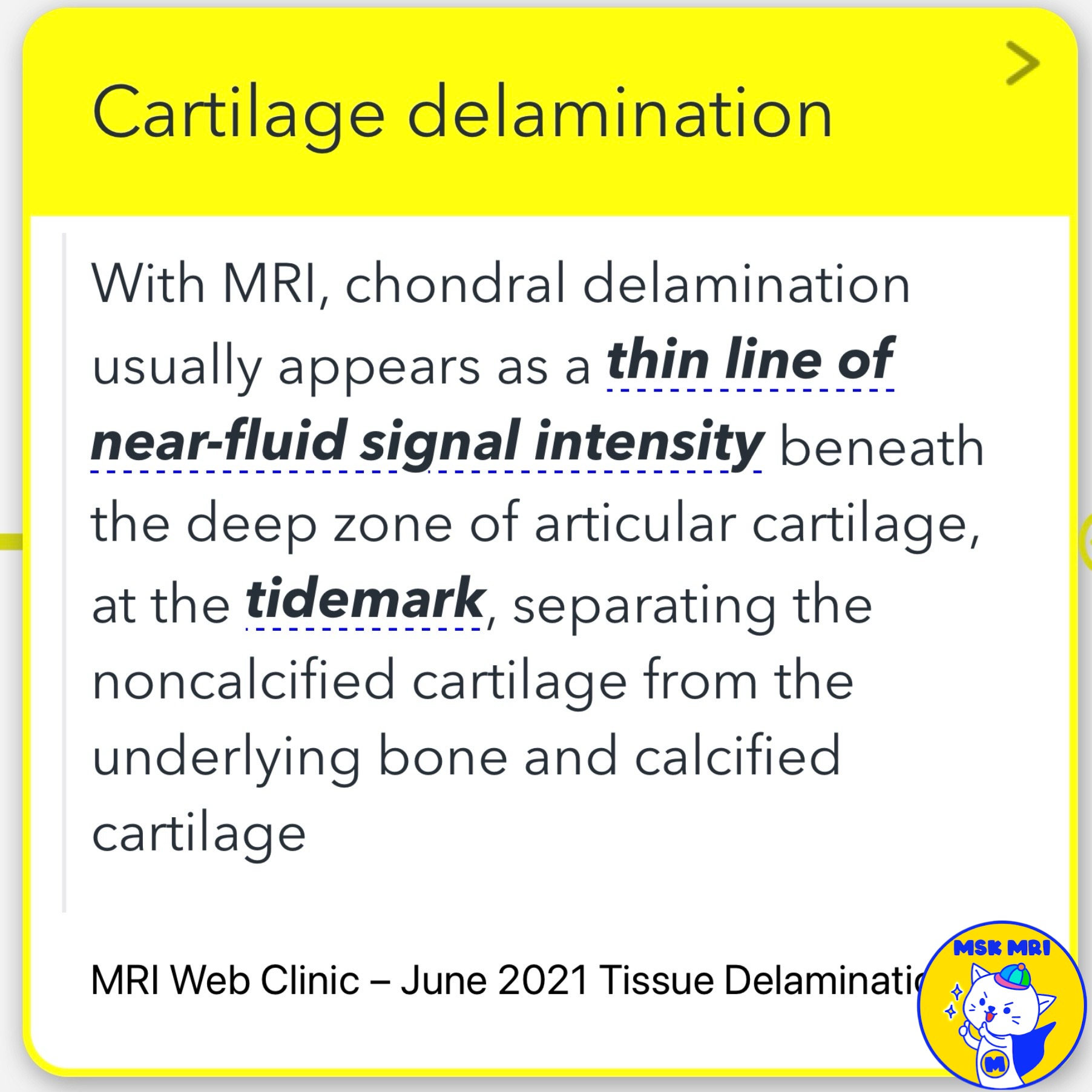
- On MRI, chondral delamination typically appears as a thin line of near-fluid signal intensity beneath the deep zone of articular cartilage at the tidemark, separating the noncalcified cartilage from the underlying bone and calcified cartilage.
- The separation zone may be thin and regular or thick and irregular, and the overlying cartilage surface may or may not be violated.
✅ Classification
- If there is superficial cartilage injury, the modified Outerbridge classification usually places delamination injuries as grade 3 (deep ulceration or a chondral flap involving 50% or more of the depth of the articular cartilage without exposure of subchondral bone) or grade 4 (exposed bone).
- According to Bauer and Jackson's arthroscopic system, delamination injuries are either flap or crater types.
- The modified ICRS classification categorizes delamination injuries as ICRS grade 3b or 3d lesions, depending on the status of the superficial cartilage.
✅ Skeletal Maturity and Delamination
- In skeletally immature individuals, the lack of well-formed tidemark and minimal calcified cartilage results in osteochondral fractures predominating over chondral injuries such as delamination.
References
- MRI Web Clinic – June 2021 Tissue Delamination
- AJR 2017; 209:W317–W321
"Visualizing MSK Radiology: A Practical Guide to Radiology Mastery"
© 2022 MSK MRI Jee Eun Lee All Rights Reserved.
No unauthorized reproduction, redistribution, or use for AI training.
#ChondralDelamination, #CartilageInjury, #Tidemark, #SubchondralBone, #MRI, #SkeletalMaturity, #TraumaticStress, #ArticularCartilage, #ICRSClassification, #Orthopedics


'✅ Knee MRI Mastery > Chap 5AB. Chondral and osteochondral' 카테고리의 다른 글
| (Fig 5-A.16) Transverse Chondral Flap Lesions in Midzone (0) | 2024.07.06 |
|---|---|
| (Fig 5-A.15) Concealed Chondral Delamination (0) | 2024.07.06 |
| (Fig 5-A.13) Fissure with Chondral Flap (0) | 2024.07.05 |
| (Fig 5-A.12) Fissure or Fissuring in Cartilage (0) | 2024.07.05 |
| (Fig 5-A.11) Hypointense Lesion in Cartilage (0) | 2024.07.05 |