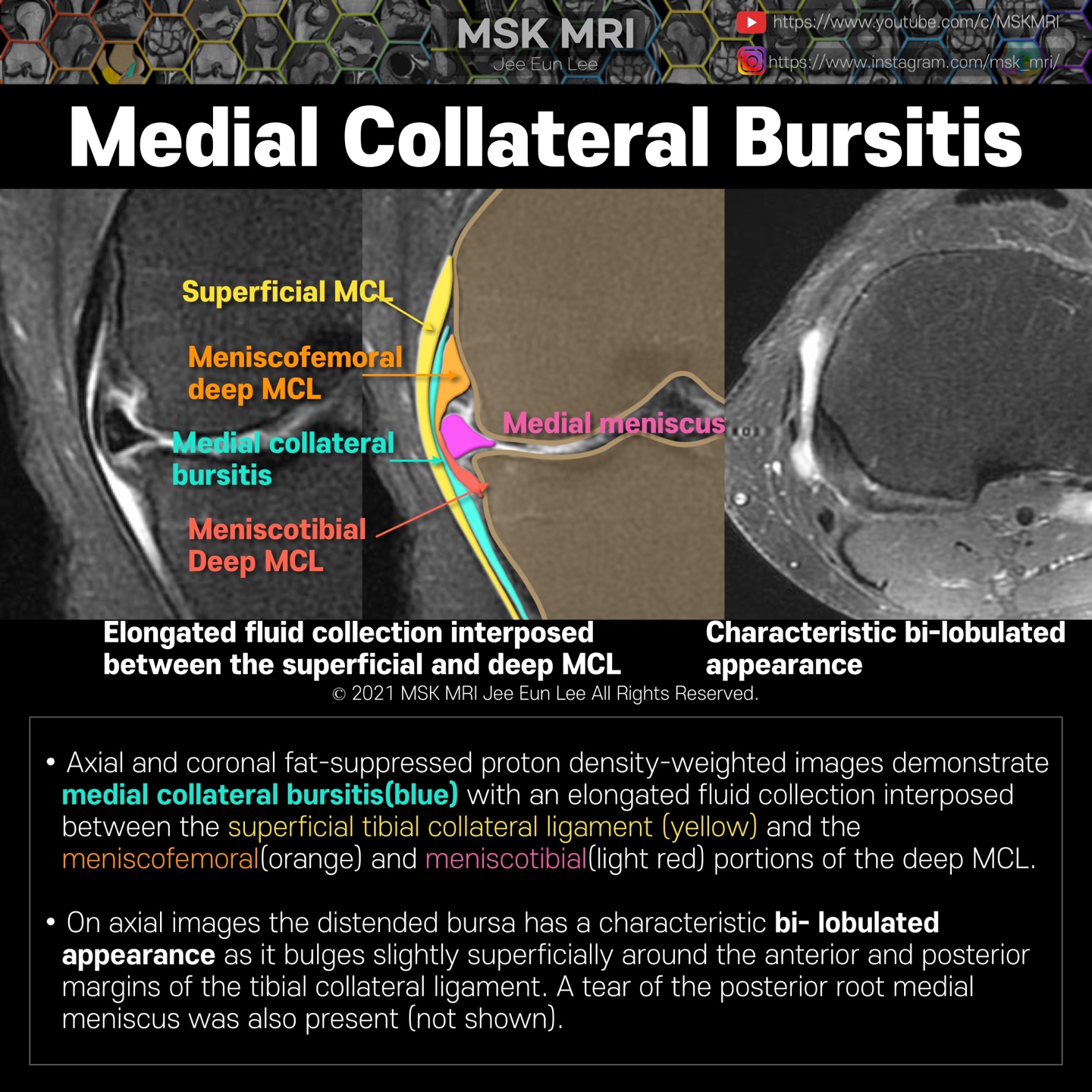
Axial and coronal fat-suppressed proton density-weighted images demonstrate medial collateral bursitis(blue) with an elongated fluid collection interposed between the superficial tibial collateral ligament (yellow) and the meniscofemoral(orange) and meniscotibial(pink) portions of the deep MCL.
On axial images, the distended bursa has a characteristic bi- lobulated appearance as it bulges slightly superficially around the anterior and posterior margins of the tibial collateral ligament.
A tear of the posterior root medial meniscus was also present (not shown).

It's not a real patient's MRI, but they are virtual images very similar to the images in the journals. The images will be created for educational purposes.
All copyrights belong to MSK MRI Jee Eun Lee.
You may not distribute or commercially exploit the content. Nor may you transmit it or store it on any other website or other forms of the electronic retrieval system.
If you would like to use an image or video for anything other than personal use, please contact me. (jamaisvu1977@gmail.com)
#Virtual MRI, #MRI illustrator, #MSKMRI © 2021 MSK MRI Jee Eun Lee All Rights Reserved.

#MSKMRI, #virtualMRI, #radiologist, #Knee_MRI, #MSKMRI_Knee, #Knee_ligament, #Knee_bursitis, #bursitis #Virtual_MRI, #MRI_illustrator


