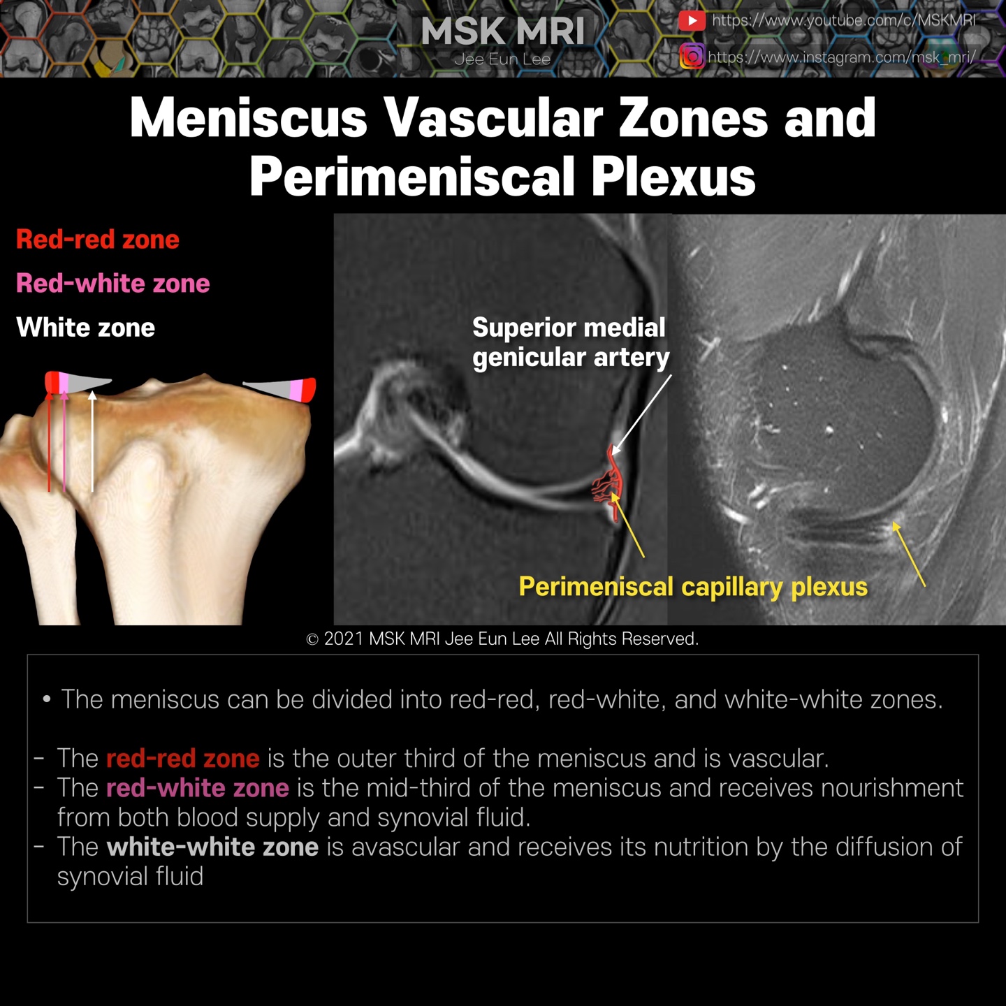
The meniscus is relatively avascular in adults, except for the peripheral 10% to 25% of the meniscus supplied by the perimeniscal capillary plexus.
Notice, on sagittal fat-suppressed PD images, this focal high signal intensity of the peripheral portion of the medial meniscus is not torn, but perimeniscal capillary plexus.
The meniscus can be divided into red-red, red-white, and white-white zones.
- The #The red-red zone is the outer third of the meniscus and the peripheral 3mm of the meniscus, maintains an excellent blood supply.
- The #red-white zone is the mid-third of the meniscus, demonstrates variable vascularity, and receives nourishment from both blood supply and synovial fluid.
- The #white-white zone extends beyond 5mm from the periphery and represents the avascular inner portion (including the free edge) of the meniscus
- The white-white zone is avascular and receives its nutrition by the diffusion of synovial fluid
It's not a real patient's MRI, but they are virtual images very similar to the images in the journals. The images will be created for educational purposes.
All copyrights belong to MSK MRI Jee Eun Lee.
You may not distribute or commercially exploit the content. Nor may you transmit it or store it on any other website or other forms of the electronic retrieval system.
If you would like to use an image or video for anything other than personal use, please contact me. (jamaisvu1977@gmail.com)
#Virtual MRI, #MRI illustrator, #MSKMRI © 2021 MSK MRI Jee Eun Lee All Rights Reserved.


#MSKMRI, #virtualMRI, #radiologist, #Knee_MRI, #MSKMRI_Knee, #Knee_anatomy, #Knee_meniscus, #meniscus, #Virtual_MRI, #MRI_illustrator,
#kneemripitfalls,


