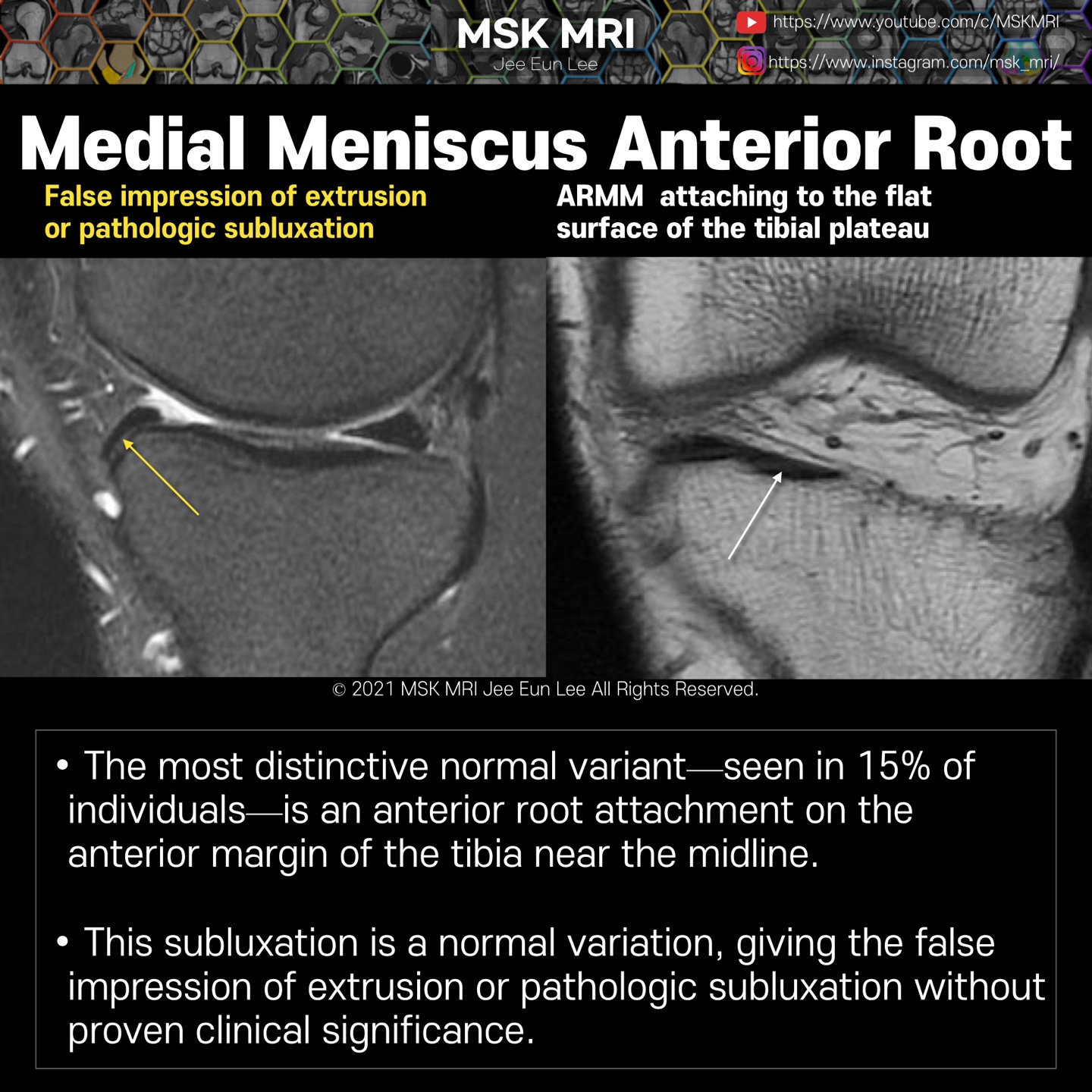
The medial meniscus becomes flattened as it transitions into the anterior root medial meniscus (ARMM)
This is made up of a single fiber bundle and is the largest of the four roots.
The anterior root lateral meniscus (ARLM) and posterior root medial meniscus (PRMM) are made up of multiple fibre bundles(consists of three or more bundles.
In 15% of individuals, the ARMM attaches onto the anterior margin of the tibia near the midline (Fig. 2c) [10], resulting in anterior subluxation of the meniscus at the mid-portion of the medial tibial plateau.
However, this is a normal variant which is of no clinical significance
This subluxation is a normal variation, giving the false impression of extrusion or pathologic subluxation
It's not a real patient's MRI, but they are virtual images very similar to the images in the journals. The images will be created for educational purposes.
All copyrights belong to MSK MRI Jee Eun Lee.
You may not distribute or commercially exploit the content. Nor may you transmit it or store it on any other website or other forms of the electronic retrieval system.
If you would like to use an image or video for anything other than personal use, please contact me. (jamaisvu1977@gmail.com)
#Virtual MRI, #MRI illustrator, #MSKMRI © 2021 MSK MRI Jee Eun Lee All Rights Reserved.


#MSKMRI, #virtualMRI, #radiologist, #Knee_MRI, #MSKMRI_Knee, #Knee_anatomy, #Knee_meniscus, #meniscus, #Virtual_MRI, #MRI_illustrator,
#Meniscusroots, #anterior_root
'Knee MRI > Meniscus' 카테고리의 다른 글
| [Anatomy_10] Medial Meniscus Posterior Root -02 (0) | 2021.09.24 |
|---|---|
| [Anatomy_09] Medial Meniscus Posterior Root -01 (0) | 2021.09.24 |
| [Anatomy_07] Attachments of the Meniscus & cruciate ligaments (0) | 2021.09.24 |
| [Anatomy_06] Meniscus Vascular Zones and Perimeniscal Plexus (0) | 2021.09.24 |
| [Anatomy_05] Normal MRI findings of medial stabilizers of the knee (0) | 2021.09.23 |


