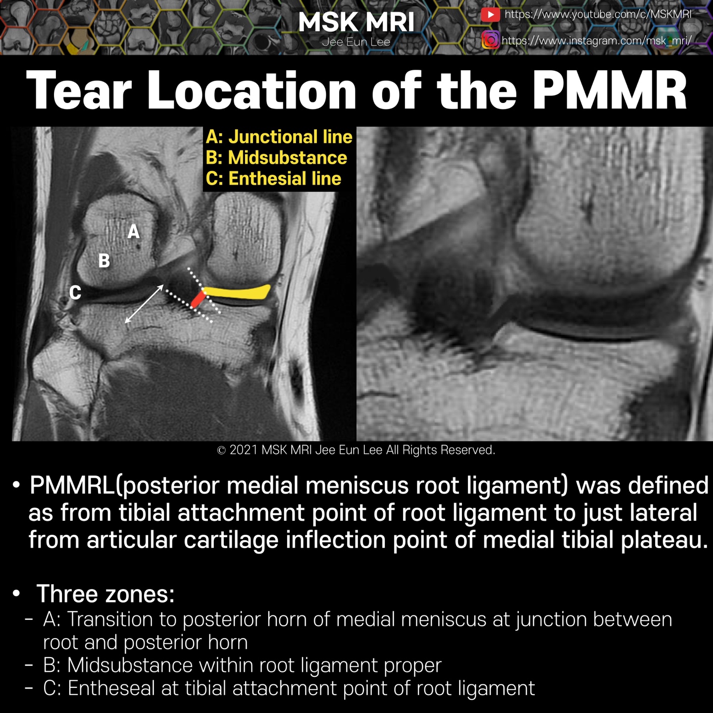
The posterior root of the medial meniscus was subcategorized into three zones: transition to posterior horn of medial meniscus at the junction between root and posterior horn (a), midsubstance within root ligament proper (b), and entheseal at the tibial attachment point of root ligament
The most common location of medial meniscal root ligament tear was junctional, between the ligament and the posterior horn, a location accounting for 93% of medial meniscal root ligament tears. Radial tears were the most common orientation, accounting for 68.3%.
Most root ligament lesions were associated with tears in the medial meniscus (86.7%)
In terms of the point of failure, we observed that the posterior root of medial meniscus(PMMRL) tears mostly occur at the junction with the PHMM followed by the midsubstance, and they least commonly manifest as an avulsion at the enthesis.
Moreover, the junction between the posterior horn and the root ligament may be structurally weaker because of the differences in morphology and collagen distribution between the fibrocartilaginous meniscus, root ligament, and enthesis
It's not a real patient's MRI, but they are virtual images very similar to the images in the journals. The images will be created for educational purposes.
All copyrights belong to MSK MRI Jee Eun Lee.
You may not distribute or commercially exploit the content. Nor may you transmit it or store it on any other website or other forms of the electronic retrieval system.
If you would like to use an image or video for anything other than personal use, please contact me. (jamaisvu1977@gmail.com)
#Virtual MRI, #MRI illustrator, #MSKMRI © 2021 MSK MRI Jee Eun Lee All Rights Reserved.


#MSKMRI, #virtualMRI, #radiologist, #Knee_MRI, #MSKMRI_Knee, #Knee_anatomy, #Knee_meniscus, #meniscus, #Virtual_MRI, #MRI_illustrator,
#Meniscusroots, #posterior_root
'Knee MRI > Meniscus' 카테고리의 다른 글
| [Anatomy_12] Normal Fissured LM Anterior Root (0) | 2021.09.24 |
|---|---|
| [Anatomy_11] Lateral Meniscus Anterior Root (0) | 2021.09.24 |
| [Anatomy_09] Medial Meniscus Posterior Root -01 (0) | 2021.09.24 |
| [Anatomy_08] Medial Meniscus Anterior Root (0) | 2021.09.24 |
| [Anatomy_07] Attachments of the Meniscus & cruciate ligaments (0) | 2021.09.24 |


