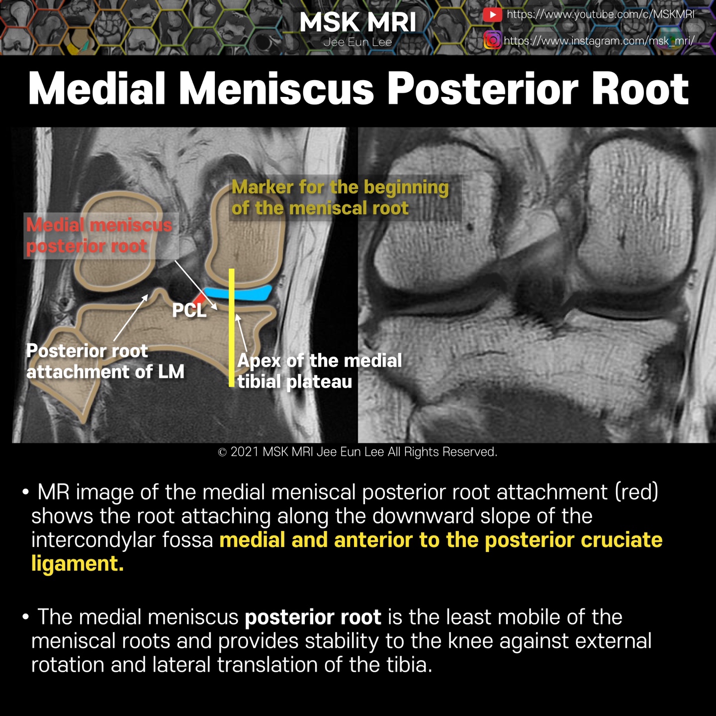
The posterior root of the medial meniscus(Red) commences just lateral to the apex of the medial tibial plateau.
The yellow line demonstrates where the cartilage of the medial tibial plateau ceases which is another marker for the beginning of the meniscal root.
The root attaches along the downward slope of the intercondylar fossa medial and anterior to the posterior cruciate ligament.
The medial meniscus posterior root is the least mobile of the meniscal roots and provides stability to the knee against external rotation and lateral translation of the tibia.
Note this demonstrates posterior root attachment of lateral meniscus.
The lateral meniscus posteriorly comes up high over the tibial spine to insert near the posterior cruciate ligament.
This upward position of the posterior horn may be the reason for the higher signal intensity of the posterior horn in all planes due to the magic angle effect.
It's not a real patient's MRI, but they are virtual images very similar to the images in the journals. The images will be created for educational purposes.
All copyrights belong to MSK MRI Jee Eun Lee.
You may not distribute or commercially exploit the content. Nor may you transmit it or store it on any other website or other forms of the electronic retrieval system.
If you would like to use an image or video for anything other than personal use, please contact me. (jamaisvu1977@gmail.com)
#Virtual MRI, #MRI illustrator, #MSKMRI © 2021 MSK MRI Jee Eun Lee All Rights Reserved.


#MSKMRI, #virtualMRI, #radiologist, #Knee_MRI, #MSKMRI_Knee, #Knee_anatomy, #Knee_meniscus, #meniscus, #Virtual_MRI, #MRI_illustrator,
#Meniscusroots, #posterior_root
'Knee MRI > Meniscus' 카테고리의 다른 글
| [Anatomy_11] Lateral Meniscus Anterior Root (0) | 2021.09.24 |
|---|---|
| [Anatomy_10] Medial Meniscus Posterior Root -02 (0) | 2021.09.24 |
| [Anatomy_08] Medial Meniscus Anterior Root (0) | 2021.09.24 |
| [Anatomy_07] Attachments of the Meniscus & cruciate ligaments (0) | 2021.09.24 |
| [Anatomy_06] Meniscus Vascular Zones and Perimeniscal Plexus (0) | 2021.09.24 |


