✨ Join the channel to enjoy the benefits!
🚀 https://www.youtube.com/channel/UC4bw7o0l2rhxn1GJZGDmT9w/join
https://youtube.com/shorts/4uXE8sluDX4
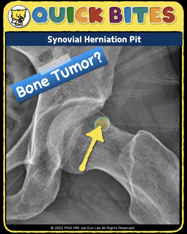
📌 Synovial Herniation Pit
✅ Definition
Synovial herniation pits, also known as Pitt pits, are common and usually incidental imaging findings, often described as fibrocystic changes at the anterosuperior femoral neck.
✅ Epidemiology
- Found in ~5% (range: 4-12%) of normal adults on X-ray.
✅ Associations
- Linked to cam morphology femoroacetabular impingement (FAI), occurring in 5-33% of cases.
- However, the causal relationship between FAI and synovial herniation pits remains unproven.
✅ Pathology
- The exact cause is debated.
- Represents a herniation of synovium or soft tissue into bone through a cortical defect.
- Commonly occurs at the anterosuperior femoral neck, but also reported in the anteroinferior region.
- Size: Typically 5 mm (range: 1-15 mm).
- More often unilateral than bilateral.
✅ Plain Radiograph / CT
- Appears as oval, round, or "8-shaped" lucencies with sclerotic margins.
- CT may reveal an overlying cortical defect.
✅ MRI
- Well-defined lesion with low peripheral signal.
- Central low T1 and high T2 signal, sometimes heterogeneous.
- Some lesions contain intralesional fat.
- Adjacent bone marrow edema is rare.
✅ Treatment & Prognosis
- Incidental finding with no clinical significance.
- Considered a “don’t touch” lesion in radiology.
- May enlarge over time but generally requires no treatment.
#SynovialHerniationPit, #PittPit, #FemoralNeck, #FAI, #HipRadiology, #MSKRadiology, #BoneLesion, #HipMRI, #SkeletalImaging, #RadiologyEducation
"Visualizing MSK Radiology: A Practical Guide to Radiology Mastery"
© 2022 MSK MRI Jee Eun Lee All Rights Reserved.
No unauthorized reproduction, redistribution, or use for AI training.