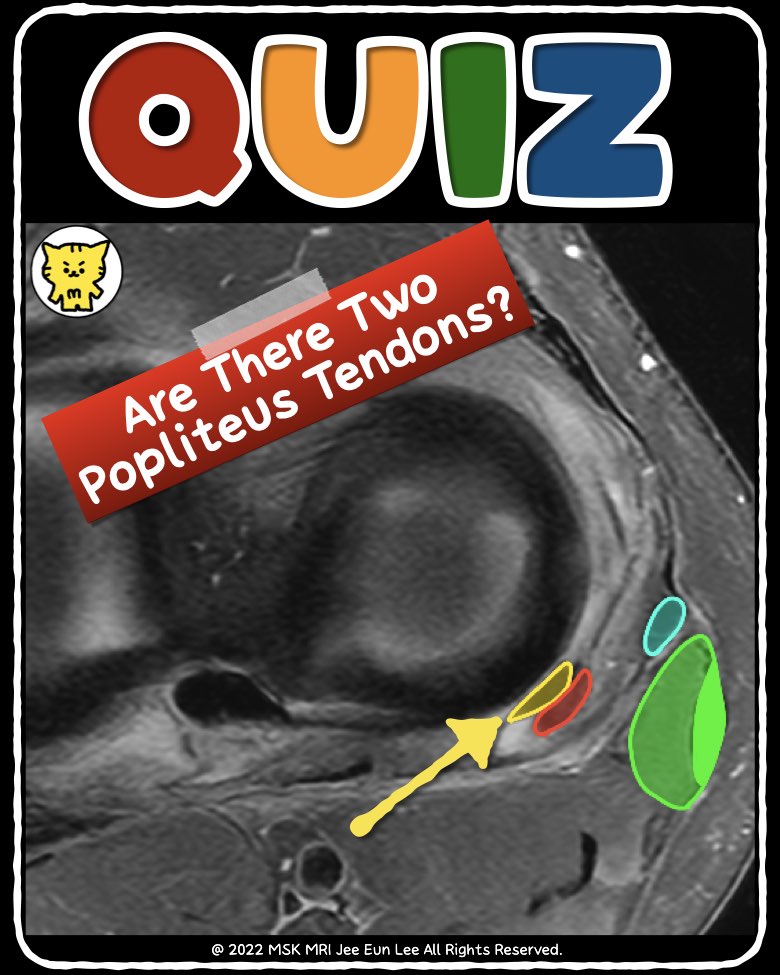
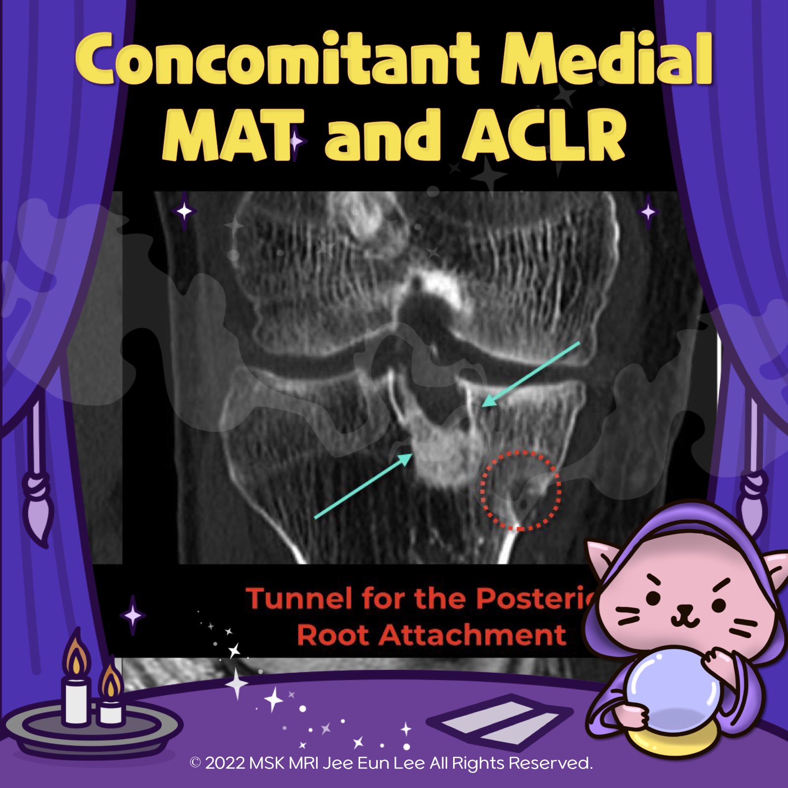
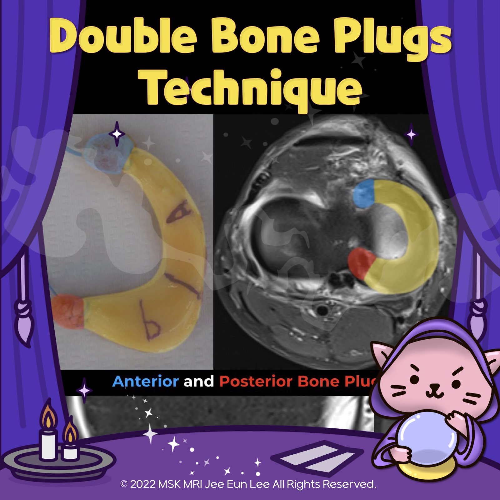
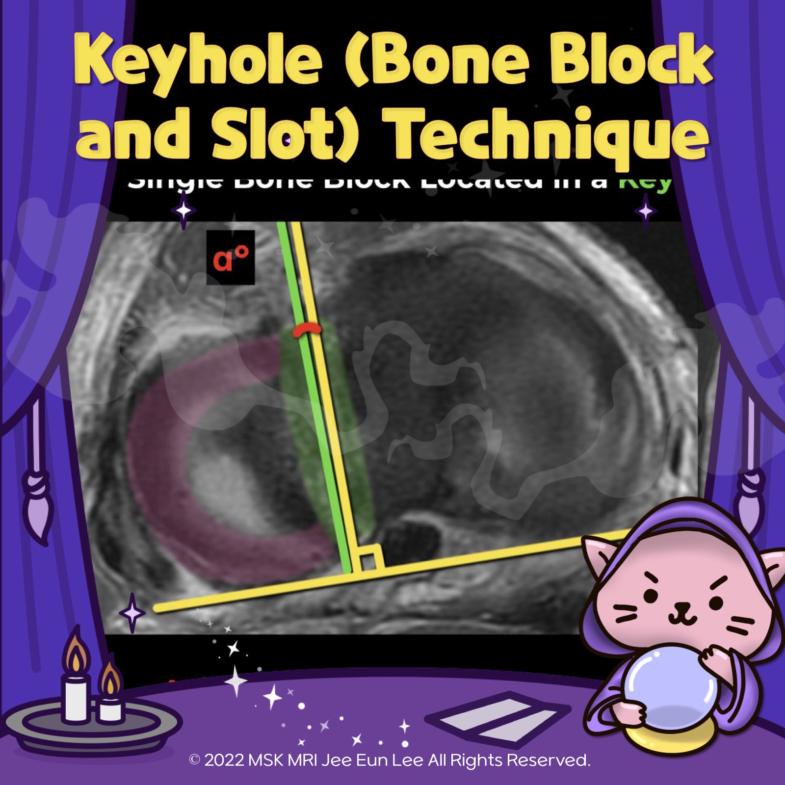
본문 바로가기

✅ Knee MRI Mastery/Chap 1. Meniscus103

(Fig 1-B.41) Type 5 Ramp Lesion https://youtu.be/kYOVoRk6YvM https://youtu.be/nKQsItyKAtM Type 5 Ramp Lesion involves two separate tears within the red-red zone of the meniscus. Similar to the 4A subtype, the meniscocapsular and meniscotibial ligaments remain intact. There are two parallel linear areas with high T2 fluid intensity signal, extending from the superior to the inferior articular surface within the red-red zone of .. 2024. 4. 7.
(Fig 1-E.23) Posterior root tear Of Meniscus Allograft Transplantation https://youtu.be/0U6wcsVeS_M https://youtu.be/SXRAadVysHc 📌 Understanding the causes of graft failure is crucial for improving meniscal allograft transplantation (MAT) outcomes. The primary reasons for graft failure include: 1️⃣ Implantation Site Issues: Failures can occur due to avulsion at the root, unsuccessful plug incorporation, or fracture of the bone bridge. 2️⃣ Healing Complications: Inc.. 2024. 2. 11.
(Fig 1-E.22) Expected Findings Of Keyhole (bone block and slot) Technique https://youtu.be/yFSeb5KicyE https://youtu.be/SXRAadVysHc 📌After Meniscal Allograft Transplantation (MAT), the allograft bone block or plugs undergo a critical process of incorporation into the host's proximal tibia, which is a significant aspect of the postoperative healing phase. Key points include: 1️⃣ Bone Incorporation: The union at the cancellous–cancellous junctions can occur as early as .. 2024. 2. 11.
(Fig 1-E.21) Concomitant MAT and ACLR and Articular cartilage restorative procedure https://youtu.be/jwtAe0gp3TY https://youtu.be/SXRAadVysHc 📌 Meniscal Allograft Transplantation (MAT) is often accompanied by additional surgical procedures to address concurrent knee issues, enhancing overall knee function and patient outcomes. Common concomitant procedures include: ➡️ ACL Reconstruction: To restore stability in patients with concurrent anterior cruciate ligament injuries. ➡️ Ar.. 2024. 2. 11.
(Fig 1-E.20) Too-small meniscal allograft and failure https://youtu.be/3KbQEifNAm8 https://youtu.be/SXRAadVysHc 📌 Preoperative Imaging and Meniscal Sizing: Precise meniscal sizing is crucial for meniscal allograft transplantation (MAT) success. A mismatch in graft size should be less than 10% to maintain the meniscus's biomechanical integrity. ✅ Specifically, too small allografts (beyond a 5-mm margin of error) can jeopardize long-term functionalit.. 2024. 2. 11.
(Fig 1-E.19) Double bone Plugs Technique for medial MAT https://youtu.be/WnUD4jPBGqA https://youtu.be/SXRAadVysHc 1️⃣ Graft Preparation: The medial meniscus graft is readied with bone plugs attached to each horn, reflecting the anatomical distance between the horns and their medial orientation relative to the ACL insertion. 2️⃣ Bony Sockets Creation: In the recipient's proximal tibia, two sockets are crafted at the sites of the original meniscal root.. 2024. 2. 11.
(Fig 1-E.18) Keyhole (bone block and slot) Technique https://youtu.be/bEa001H5rKA https://youtu.be/SXRAadVysHc 1️⃣ Meniscal Extrusion: It's a common occurrence post-MAT, yet there's no universally accepted standard for what constitutes normal meniscal extrusion. The link between clinical outcomes and radiologic findings regarding extrusion remains unclear, indicating a need for further study to understand its implications fully. 2️⃣ Axial Trough A.. 2024. 2. 11.
(Fig 1-E.17) Complete Retear of Transtibial Pullout Repair failure https://youtu.be/peGP49QExi8 https://youtu.be/SXRAadVysHc ✅Summary of Meniscal Root Repair Healing Criteria and MRI Interpretation Challenges • Complete Healing: Defined as the continuity of the repaired meniscal root across axial, coronal, and sagittal planes. • Partial Healing: Identified when continuity is observed in one or two of these planes. • Repeat Tear: Recognized by the absence of con.. 2024. 2. 10.
(Fig 1-E.16) Partial healing, a partial tear of Transtibial Pullout Repair https://youtu.be/zQJLfC2g2ZY https://youtu.be/SXRAadVysHc 1️⃣ Summary of Meniscal Root Repair Healing Criteria and MRI Interpretation Challenges • Complete Healing: Defined as the continuity of the repaired meniscal root across axial, coronal, and sagittal planes. • Partial Healing: Identified when continuity is observed in one or two of these planes. • Repeat Tear: Recognized by the absence of .. 2024. 2. 10.
(Fig 1-E.15) Transtibial Pullout Repair of Lateral Meniscus Posterior Root Tear https://youtu.be/-sLDIAWVAsE https://youtu.be/SXRAadVysHc ★ Transtibial Pullout Repair of Lateral Meniscus Posterior Root Tear ★ The lateral root repair technique is crucial, especially when dealing with lateral root tears that often accompany anterior cruciate ligament (ACL) injuries. The procedure involves several key steps: 1️⃣ Tissue Preparation and Suture Passage: These initial steps are ca.. 2024. 2. 10.
(Fig 1-E.13) Transtibial Pullout Repair https://youtu.be/H08xEv_X1cE https://youtu.be/SXRAadVysHc 📌Transtibial Pullout Repair 📌 • Detachment of Posterior Root: A detached posterior root of the meniscus is functionally equivalent to a total meniscectomy, leading to a significant loss in the meniscus’s ability to withstand hoop stress. This condition severely compromises the knee’s structural integrity and function. • Association with O.. 2024. 2. 10.
(Fig 1-E.12) Recurrent Tear in meniscal repair -4 https://youtu.be/nylyCmV7n3I https://youtu.be/SXRAadVysHc 📌Direct Signs of Recurrent Tear in Postoperative Meniscus ① Increased signal nearly that of the joint fluid contacting the articular surface ② Change in the signal intensity pattern through the meniscus ③ Tear at another location: New meniscal tear pattern ④ Displaced flap: fragmentation of meniscal remnant 📌Indirect Signs of Recurrent Te.. 2024. 2. 10.
(Fig 1-E.11) Recurrent Tear in meniscal repair -3 https://youtu.be/2Di69B0Uwh0 https://youtu.be/SXRAadVysHc 📌 Suture Tracks in Meniscal Tissue Identification: Suture tracks within the meniscal tissue are discernible across all MRI pulse sequences. However, their visibility is notably enhanced in PD (Proton Density) Fat Suppressed (PDFS) sequences. ➡️ Suture Tracks Appearance: Short Axis: Manifest as dot-like images, showcasing an intermediate t.. 2024. 2. 10.
(Fig 1-E.10) Recurrent Tear in meniscal repair -2 https://youtu.be/MiCe5A-Nv7k https://youtu.be/SXRAadVysHc ★ Recurrent Tear in Meniscal Repair from MR Imaging ★ 📌 Specific Indicator: A fluid-like high T2-weighted signal over 3 months post-surgery specifically indicates a meniscal tear, though its sensitivity is limited. 📌 Enhanced Accuracy: Additional MR imaging criteria—such as non-concordant abnormal meniscal morphology post-surgery, displac.. 2024. 2. 10.
(Fig 1-E.09) Recurrent Tear in meniscal repair -1 https://youtu.be/efTV1COwS5Q https://youtu.be/SXRAadVysHc 🌟Failed Meniscal Repair 🌟 1️⃣ Signal intensity Definitively surfacing T2 high signal (such as fluid signal) after at least three months following the initial repair. Sensitivity and specificity of approximately 60% and 90% 2️⃣ Abnormal meniscal morphology Inconsistencies with prior surgery, a displaced flap, or a tear at a different locat.. 2024. 2. 10.
(Fig 1-E.08) Expected appearance of meniscal repair -3 https://youtu.be/deyXAY6Kz4Q https://youtu.be/SXRAadVysHc 📌 All-inside meniscal repair technique The all-inside meniscal repair technique often results in a grade 3 intensity on MRI scans postoperatively, making it challenging to distinguish between healing menisci and repeat tears. This similarity limits the accuracy of MR imaging for postoperative assessments. Observations indicate that MRI im.. 2024. 2. 9.
(Fig 1-E.07) Expected appearance of meniscal repair -2 https://youtu.be/GI2cd3D99o4 https://youtu.be/SXRAadVysHc 📌Normal MRI appearance after meniscal repair In conventional MR imaging following meniscal repair, a hyperintense signal at the repair site can persist for over 10 years post-surgery. Importantly, even full-thickness signal abnormalities do not reliably indicate recurrent tears. This persistent hyperintense signal could result from either.. 2024. 2. 9.
(Fig 1-E.06) Expected appearance of meniscal repair -1 https://youtu.be/rwUOEnkNw6g https://youtu.be/SXRAadVysHc 📌 Expected appearance of meniscal repair After a meniscal repair without any resection, the postoperative morphology of the meniscus appears essentially normal on MRI, contrasting with the imaging seen after partial meniscectomy. A meniscus is considered healed if no fluid signal is detected in the area of repair. "Visualizing MSK Radiolo.. 2024. 2. 9.
(Fig 1-E.05) Recurrent Tear in partial meniscectomy -2 https://youtu.be/zLeVN2PQLLI https://youtu.be/SXRAadVysHc 🧐Direct Signs of Recurrent Tear in Postoperative Meniscus ① Increased signal nearly that of the joint fluid contacting the articular surface ② Change in the signal intensity pattern through the meniscus ③ Tear at another location: New meniscal tear pattern ④ Displaced flap: fragmentation of meniscal remnant "Visualizing MSK Radiology: A P.. 2024. 2. 9.
(Fig 1-E.04) Recurrent Tear in partial meniscectomy -1 https://youtu.be/1hdvwRZz86g https://youtu.be/SXRAadVysHc 📌Direct Signs of Recurrent Tear in Postoperative Meniscus ① Increased signal nearly that of the joint fluid contacting the articular surface ② Change in the signal intensity pattern through the meniscus ③ Tear at another location: New meniscal tear pattern ④ Displaced flap: fragmentation of meniscal remnant 📌Indirect Signs of Recurrent Te.. 2024. 2. 9.
(Fig 1-E.03) Expected appearance of High-grade partial meniscectomy https://youtu.be/NzeljQPtQtU https://youtu.be/SXRAadVysHc ✅ High-grade partial meniscectomy (>25%) In cases of high-grade partial meniscectomy, where more than 25% of the meniscus is removed, diagnosing tears becomes less accurate. For T2-weighted MRI scans, a high signal intensity specifically indicates synovial fluid within a tear's cleft with over 90% specificity, but this sign lacks sensitiv.. 2024. 2. 9.