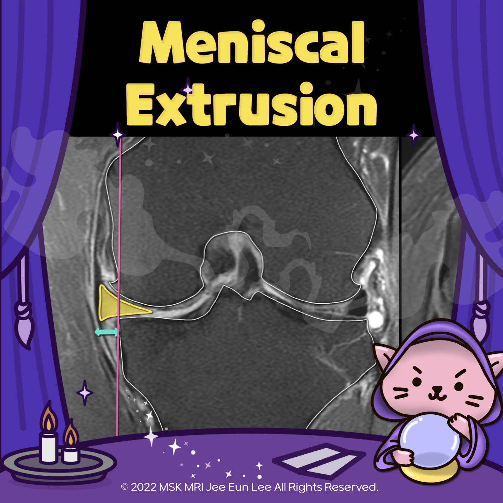
본문 바로가기

✅ Knee MRI Mastery/Chap 1. Meniscus103

(Fig 1-B.27) Giraffe neck sign of root tear https://youtu.be/bNgBZdQCOSs https://youtube.com/shorts/a0GgvplMLQ4 https://youtu.be/nKQsItyKAtM Giraffe Neck Sign The "Giraffe Neck Sign" refers to a trapezoidal shape of the meniscal body, characterized by a thin inner margin and a larger outer margin. This sign has been recently identified as an indicator of medial meniscus posterior root tears. "Visualizing MSK Radiology: A Practical Guide t.. 2024. 2. 6.
(Fig 1-B.26) Direct signs of root tears https://youtu.be/w113IDroK1s https://youtu.be/nKQsItyKAtM Both primary and secondary MRI findings can aid in the diagnosis of posterior root tears. Among the primary MRI signs of root tears, three commonly used findings are: Sagittal Ghost Sign: This sign is characterized by a hyperintense white signal intensity that leads to the disappearance or abrupt blunting of the normal low-signal-intensit.. 2024. 2. 6.
(Fig 1-B.25) MRI Classification of Posterior Medial Meniscal Root Injuries https://youtu.be/eaqz6AnS_sE https://youtube.com/shorts/zwGm1C5oAEI https://youtube.com/shorts/6ygFdDCmIMM https://youtu.be/nKQsItyKAtM MRI Classification of Posterior Medial Meniscal Root Injuries 1. Root Degeneration: Characterized by thickening of the root with intrasubstance hyperintensity that does not reach the articular surface. 2. Root Partial Tear: Involves abnormal signal intensity ext.. 2024. 2. 6.
(Fig 1-B.24) bucket-handle tear versus Hemi-Bucket handle tear https://youtu.be/IpQCx8TqpBc https://youtu.be/nKQsItyKAtM Hemi-bucket-handle tears "A hemi-bucket-handle tear is an undersurface flap tear of the medial meniscus, where meniscal tissue is displaced toward the intercondylar notch. (there is no truncation of the free edge of the medial meniscus; instead, there is an undersurface flap tear with a predominantly horizontal orientation.) Surgeons are .. 2024. 2. 6.
(Fig 1-B.23) Broken Bucket Handle Meniscal Tears https://youtu.be/x4bLWBfxtfs https://youtu.be/nKQsItyKAtM Broken bucket handle meniscal tears This entity refers to a bucket handle tear with an additional rupture of the meniscal fragment. ​​Broken bucket handle tears are often difficult to identify on MRI. However, it is a crucial injury to be aware of and recognize, as the mobility of the ruptured fragments results in significant pain and joi.. 2024. 2. 5.
(Fig 1-B.22) Absent Bow Tie and Disproportional Posterior Horn Signs https://youtu.be/KI-DwuO7jCY https://youtube.com/shorts/tiexOL6323Q https://youtu.be/nKQsItyKAtM Absent bow tie sign The absent bow tie sign, where the innermost bow tie is not present, can also be seen in a small or pediatric patient and with a radial tear of the body, macerated meniscus, or prior partial meniscectomy Disproportionally small posterior horn The disproportional posterior horn sig.. 2024. 2. 4.
(Fig 1-B.22) Absent Bow Tie and Disproportional Posterior Horn Signs https://youtu.be/TT-PykByPOQ https://youtu.be/nKQsItyKAtM Absent bow tie sign The absent bow tie sign, where the innermost bow tie is not present, can also be seen in a small or pediatric patient and with a radial tear of the body, macerated meniscus, or prior partial meniscectomy Disproportionally small posterior horn The disproportional posterior horn sign implies the presence of a larger meni.. 2024. 2. 4.
(Fig 1-B.21) Double ACL Sign Bucket-Handle Tear (Lateral meniscus) https://youtu.be/OHke0RUhmi0 https://youtube.com/shorts/mylJNLBdp1Q https://youtu.be/nKQsItyKAtM Double ACL Sign: Occasionally, a bucket-handle tear of the lateral meniscus can present with what's known as the "Double ACL Sign." This sign occurs when the torn fragment is positioned just posterior to the anterior cruciate ligament (ACL). It's important to note that this sign is specific to latera.. 2024. 2. 3.
(Fig 1-B.20) Double PCL Sign Bucket-Handle Tear (Medial meniscus) https://youtu.be/ozN1U6WaN4s https://youtube.com/shorts/ktPsagkexiE https://youtu.be/nKQsItyKAtM 🧐 Double Posterior Cruciate Ligament (PCL) Sign: Double posterior cruciate ligament (PCL) sign results from meniscal material in the notch, inferior and parallel to the PCL in the same sagittal plane. The intact ACL serves as a barrier, preventing further lateral displacement of the meniscal segment... 2024. 2. 2.
(Fig 1-B.19) Complex meniscal tear https://youtu.be/rwTxz7w45Xs https://youtube.com/shorts/58SqVlZQgAQ https://youtu.be/nKQsItyKAtM 🔍 Understanding Complex Meniscal Tears A combination of radial, horizontal, and longitudinal components. Extends on multiple planes, possibly forming separate flaps. 📸 On MRI: Look for vertical & horizontal patterns, hinting at a fragmented structure. 🚨 'Complex Tear': Indicates extensive distortion .. 2024. 2. 1.
(Fig 1-B.18) Change in the slope of a flap tear https://youtu.be/F0VOgHL4EYg https://youtube.com/shorts/05xM7BZFR_8 https://youtu.be/nKQsItyKAtM 🦵 Recognizing Flap Tears in Meniscus 🦵 When dealing with meniscal flap tears, keep an eye out for a crucial indicator – a change in the slope of the meniscus's superior surface. This change signals a shift in the tear's direction, forming the characteristic flap. 🔍 Changing Slope Sign: Flap tears oft.. 2024. 1. 31.
(Fig 1-B.17) Horizontal tear versus horizontal flap tear https://youtu.be/RvrGR990D0Q https://youtube.com/shorts/ATQAm2YVYSU https://youtu.be/nKQsItyKAtM 🦵 Horizontal Flap Tear vs. Horizontal Tear 🦵 Horizontal Flap Tear In a horizontal flap tear, we observe a meniscal fragment displaced deeper than the Medial Collateral Ligament (MCL). However, in a horizontal cleavage tear, there is no meniscal fragment situated deeper than the MCL. 👁️‍🗨️ Diagnosing .. 2024. 1. 30.
(Fig 1-B.16) Tips for detecting flap tear in MRI findings https://youtu.be/Xj1xqcs9RSo https://youtube.com/shorts/lLrrwrie-wE https://youtu.be/nKQsItyKAtM 🦵Radial Tear: 🔄 The meniscus-free edge is blunted, suggesting a partial thickness tear. 🔍 The radial tear exhibits a cleft with vertical high signal intensity. 🚫 However, there is no relative deficiency of the inferior surface of the meniscus. 🦵Flap Tear: Characteristic findings in flap tears include.. 2024. 1. 29.
(Fig 1-B.15) Displaced flap tear into the Popliteal hiatus https://youtu.be/A0HbFl2NEkE https://youtube.com/shorts/KHtzDrMSN64 https://youtu.be/nKQsItyKAtM 🧲 MRI Insights: Displaced Flap Tear into the Popliteal Hiatus 🏥 🌀 Beyond a Simple Tear: This case isn't just a full-thickness radial tear. It's a displaced flap tear, with a trajectory moving posteromedially due to a tear in the body of the lateral meniscus. Originating from the lateral meniscus's bo.. 2024. 1. 28.
(Fig 1-B.14) displaced flap tear into the intercondylar notch -2 https://youtu.be/vfRwbrm5l3M https://youtube.com/shorts/6Ws0F1EzU2g https://youtube.com/shorts/IodGjmAGzgA https://youtu.be/nKQsItyKAtM Flap Tear vs. Bucket-Handle Tear: Flap tears, unlike bucket-handle tears, do not show complete posterior-to-anterior extension and displacement of the involved meniscal fragment. In this scenario, the flap tear mimics a bucket-handle tear, yet distinct differenc.. 2024. 1. 27.
(Fig 1-B.13) Displaced flap tear into the intercondylar notch -1 https://youtu.be/sMbeDDeLTXs https://youtu.be/nKQsItyKAtM 🔍 Exploring Displaced Meniscal Tears in MRI Understanding meniscal tears is key in knee MRI. Let’s break down the three main types of tears involving displaced meniscal material: 1️⃣ Bucket-Handle Tear with Displacement 2️⃣ Flap Tear with Displacement 3️⃣ Free Fragment Displacement ⚠️ Why It Matters: Identifying any displaced fragments is.. 2024. 1. 26.
(Fig 1-B.11) Horizontal flap tears -2 Superior recess https://youtu.be/fy92wt6sc88 https://youtu.be/nKQsItyKAtM Insight into Horizontal Flap Tears of the Meniscus Patterns of Displacement: For medial meniscus tears, 93% of fragments are located medially or posterolaterally, commonly in the superior or inferior recesses. In contrast, lateral meniscal fragments exhibit a more varied distribution. In medial cases, two-thirds show the fragment displace.. 2024. 1. 25.
(Fig 1-B.10) Horizontal flap tears -1, Meniscotibial recess https://youtu.be/CCFU0yNkwoU https://youtu.be/nKQsItyKAtM Insight into Horizontal Flap Tears of the Meniscus What Are Horizontal Flap Tears? Horizontal flap tears involve a short segment of the meniscus with the displacement of a meniscal leaf. These tears commonly result in the displacement of the meniscal fragment, usually found in the posterior intercondylar notch or the gutters. The medial m.. 2024. 1. 24.
(Fig 1-B.09) Vertical flap tears https://youtu.be/PjQ5Dk5ub4k https://youtu.be/nKQsItyKAtM Vertical Flap Tear, also known colloquially as the Parrot Beak Tear. This particular form of tear is notable for its unique composition and the resulting displacement pattern. Composition of the Tear: The Vertical Flap Tear is characterized by having both radial and longitudinal components. Origin and Progression: Classically, this injury.. 2024. 1. 23.
(Fig 1-B.08) Marching cleft sign in a radial tear https://youtu.be/QM6RshHhemE https://youtu.be/nKQsItyKAtM Radial tears can occur in various parts of the meniscus, including the anterior/posterior horns or the body. The manifestation of these tears on MRI varies based on the tear's thickness and location. Full Thickness Radial Tear - The Ghost Meniscus Sign: Partial Thickness Radial Tear - The Truncated Triangle Sign: The Cleft Sign in Radial .. 2024. 1. 22.
(Fig 1-B.07) Full-thickness Radial Meniscal Tears https://youtu.be/a8ghran53aw https://youtu.be/nKQsItyKAtM 📌 Significance of Radial Tears: • Unlike horizontal and longitudinal tears, radial tears significantly disrupt meniscal hoop strength. • The deeper the tear, the more it impacts the biomechanical function of the menisci. 🔬 Radiologic Signs to Detect Radial Tears: 1. Truncated Triangle Sign: • Truncation of the free edge, usually indicatin.. 2024. 1. 21.