★ Ganglia vs. Parameniscal Cysts ★
1️⃣ Parameniscal Cyst
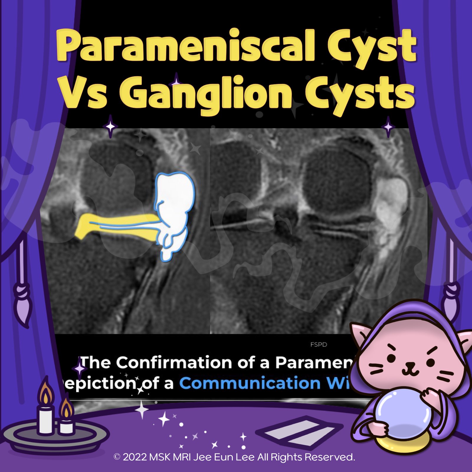
- Can appear similar to a ganglion but is usually smaller.
- Typically associated with an underlying meniscal tear.
- Diagnosis confirmed by showing a connection with an adjacent meniscal tear on MRI, regardless of the pulse sequence used.
2️⃣ Infrapatellar Ganglion
✅ Characteristics
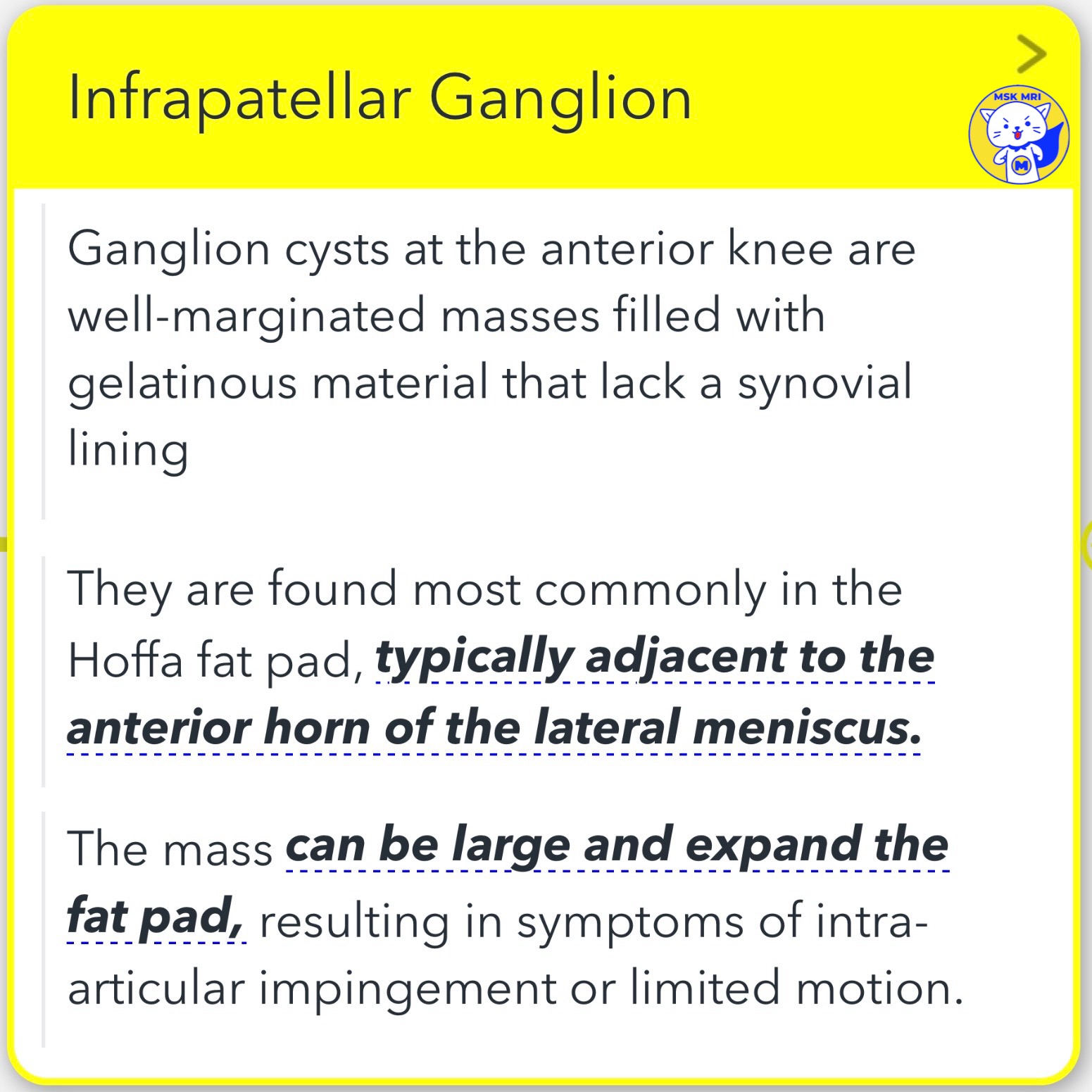
- Well-marginated masses located at the anterior knee.
- Filled with gelatinous material and lack a synovial lining.
- Most commonly found in the Hoffa fat pad, adjacent to the anterior horn of the lateral meniscus.
✅ Clinical Presentation
- The mass may be large, expanding the fat pad.
- Symptoms may include intra-articular impingement or limited motion.
✅ MRI Appearance
- Appears as a well-defined, homogeneous, non-enhancing fluid-filled collection.
- Rupture or leakage can cause ill-defined adjacent edema.
"Visualizing MSK Radiology: A Practical Guide to Radiology Mastery"
© 2022 MSK MRI Jee Eun Lee All Rights Reserved.
#VisualizingMSK #meniscaltear #meniscus #ganglioncyst


'✅ Knee MRI Mastery > Chap 1. Meniscus' 카테고리의 다른 글
| (Fig 1-C.15) Lateral Meniscal Extrusion (0) | 2024.02.08 |
|---|---|
| (Fig 1-C.14) Medial Meniscal Extrusion (0) | 2024.02.08 |
| (Fig 1-C.12) Meniscal Cysts of lateral meniscus (0) | 2024.02.08 |
| (Fig 1-C.11) Meniscal Cysts of medial meniscus (0) | 2024.02.08 |
| (Fig 1-C.10) Peripheral Meniscal Instability (0) | 2024.02.08 |