★ Meniscal Cyst Overview: ★
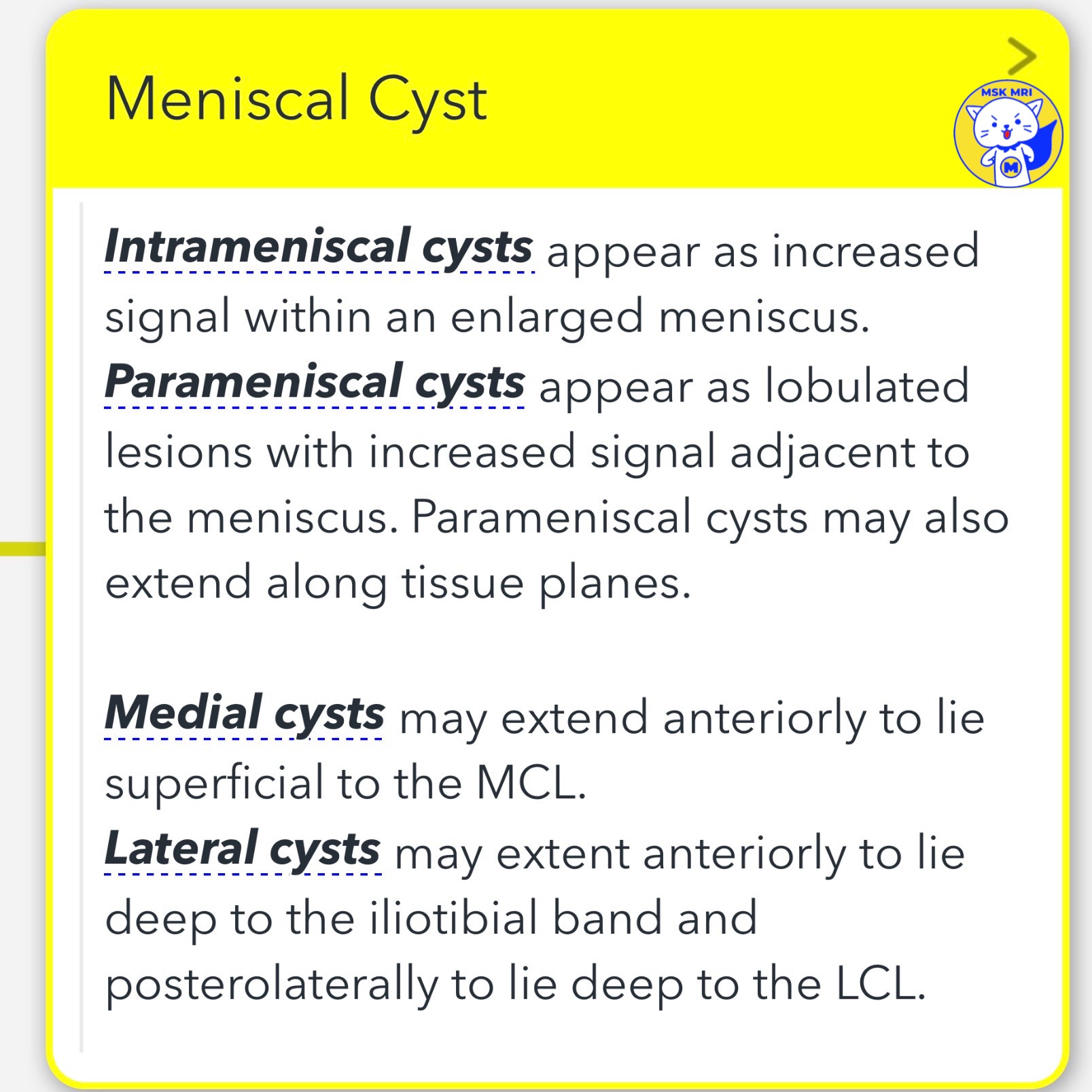
1️⃣ Intrameniscal Cysts:
- Appear as an increased signal within an enlarged meniscus.
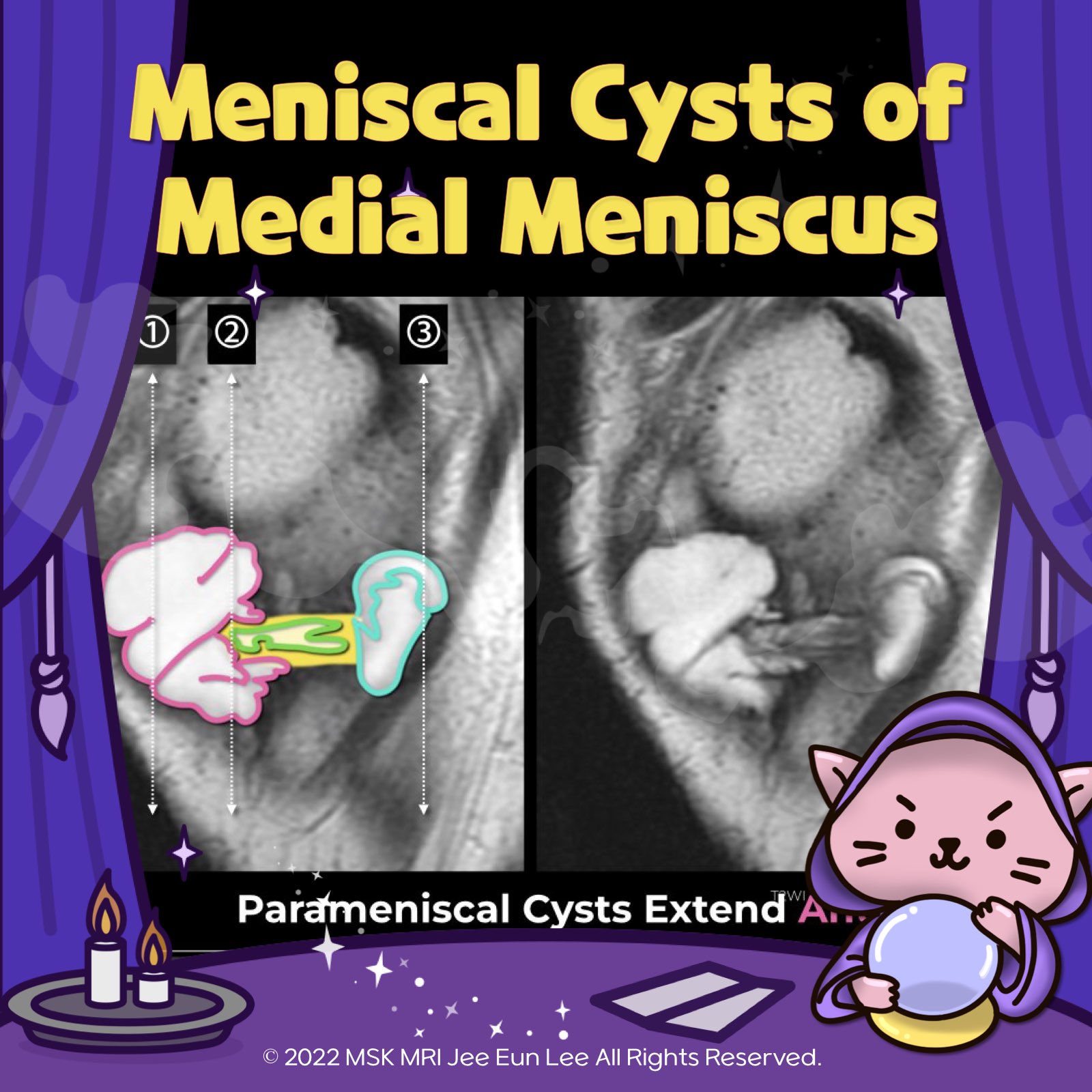
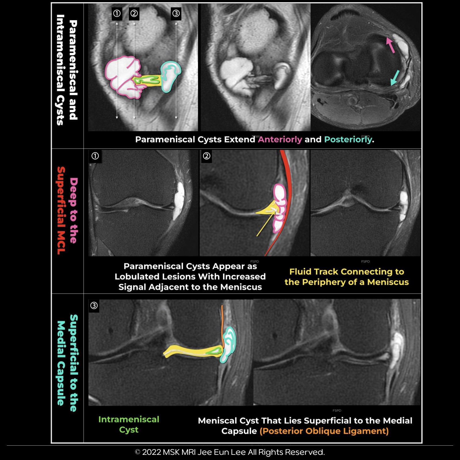
2️⃣ Parameniscal Cysts:
- Appearance: Lobulated lesions with increased signal adjacent to the meniscus.
- Location: May extend along tissue planes.
✅ Medial Cysts:
- Can extend anteriorly to lie superficial to the Medial Collateral Ligament (MCL).
✅ Lateral Cysts:
- May extend anteriorly to lie deep to the iliotibial band and posterolaterally to lie deep to the Lateral Collateral Ligament (LCL).
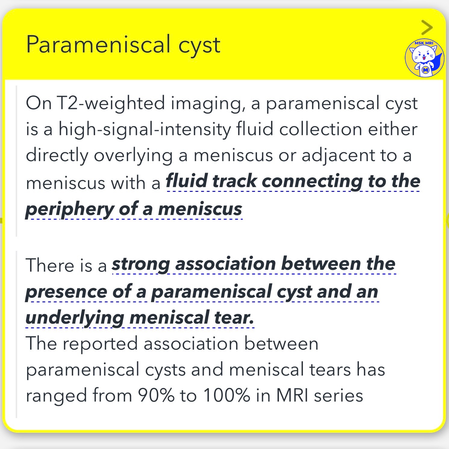
3️⃣ Parameniscal Cyst Specifics on T2-weighted Imaging:
- It is defined as a high-signal-intensity fluid collection either directly overlying a meniscus or adjacent to a meniscus with a fluid track connecting to the periphery of a meniscus.
4️⃣ Association with Meniscal Tears:
- There is a strong association between the presence of a parameniscal cyst and an underlying meniscal tear.
- The association rates between parameniscal cysts and meniscal tears in MRI series have ranged from 90% to 100%.
- 📌 Exception: At the anterior horn of the lateral meniscus, an underlying meniscal tear was found in only 64% of patients with these parameniscal cysts.
"Visualizing MSK Radiology: A Practical Guide to Radiology Mastery"
© 2022 MSK MRI Jee Eun Lee All Rights Reserved.
#VisualizingMSK #meniscaltear #meniscus
'✅ Knee MRI Mastery > Chap 1. Meniscus' 카테고리의 다른 글
| (Fig 1-C.13) Parameniscal cyst versus ganglion cysts (0) | 2024.02.08 |
|---|---|
| (Fig 1-C.12) Meniscal Cysts of lateral meniscus (0) | 2024.02.08 |
| (Fig 1-C.10) Peripheral Meniscal Instability (0) | 2024.02.08 |
| (Fig 1-C.08) Degenerated and torn lateral discoid meniscus (0) | 2024.02.08 |
| (Fig 1-C.07) Meniscal shape deformation and torn lateral discoid meniscus (1) | 2024.02.08 |