★ Meniscal Cysts Overview ★
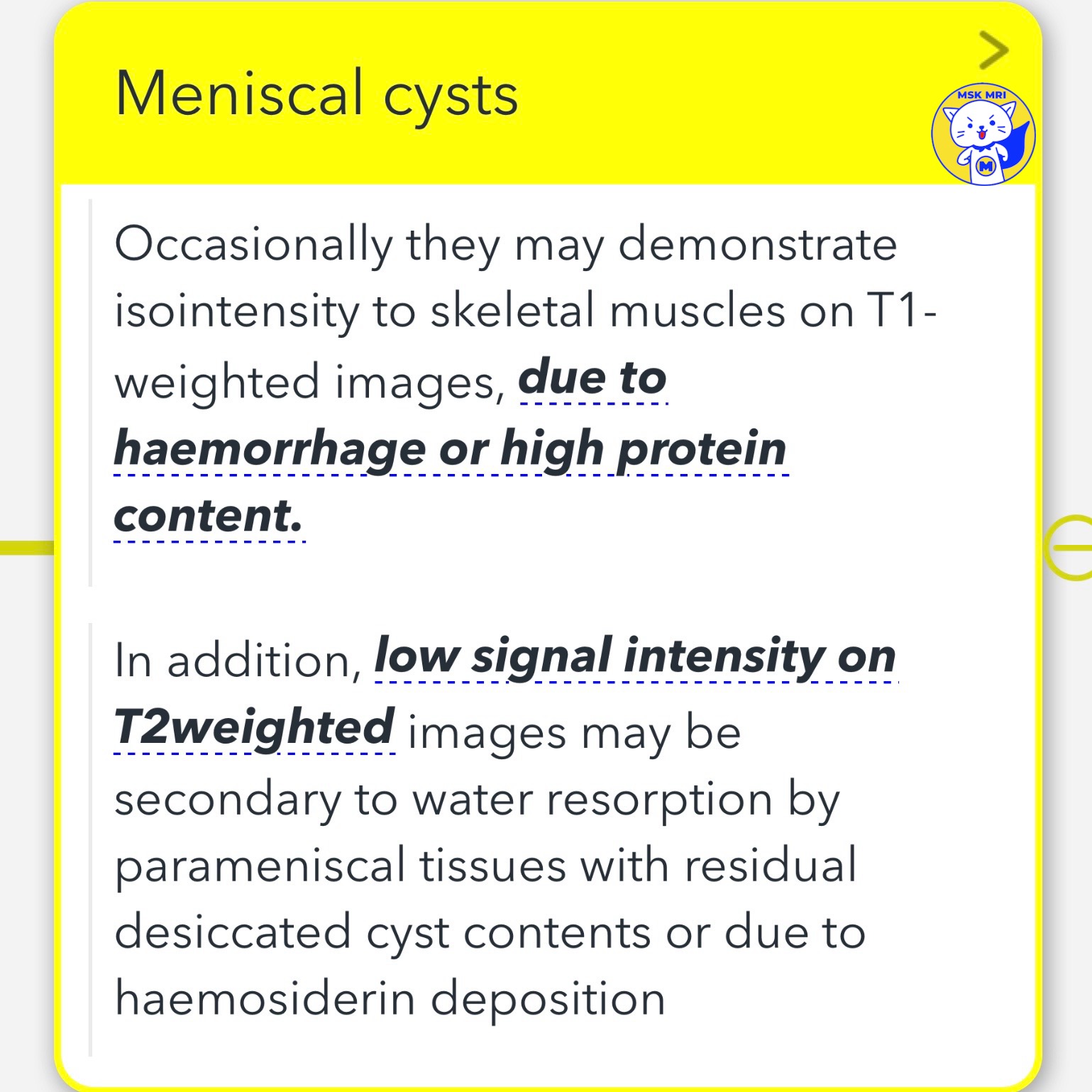
- Meniscal cysts can sometimes show the same signal intensity as skeletal muscles on T1-weighted MRI images.
- This similarity can be attributed to the presence of hemorrhage or high protein content within the cysts.
- On T2-weighted images, these cysts may appear with low signal intensity, which could be due to the absorption of water by surrounding tissues, leaving behind desiccated cyst contents, or the deposition of hemosiderin.
1️⃣ Medial Meniscal (MM) Parameniscal Cysts
✅ Common Location: Adjacent to the meniscus posterior horn, reflecting the higher incidence of tears in this area.
✅ Key Consideration: Medial parameniscal cysts can extend through the joint capsule's soft tissues or the medial collateral ligament (MCL), appearing distant from their original tear site.
2️⃣ Lateral Meniscal (LM) Parameniscal Cysts
✅ Common Location: Typically found near the anterior horn or the body of the lateral meniscus.
✅ Mobility and Extension: Due to the lateral meniscus's loose attachment to the joint capsule, anterior horn and body cysts can penetrate lateral structures and reach areas deep to the iliotibial tract.
✅ Underlying Tears: In 36% of cases, meniscal tears were not detected in patients with cysts located near the anterior horn or the anterior horn-body regions of the lateral meniscus.
✅ Patterns of Anterior Cysts: These can be located directly in front of the meniscus, dissect into the anterior root, or extend into the Hoffa's fat pad.
"Visualizing MSK Radiology: A Practical Guide to Radiology Mastery"
© 2022 MSK MRI Jee Eun Lee All Rights Reserved.
#VisualizingMSK #meniscaltear #meniscus


'✅ Knee MRI Mastery > Chap 1. Meniscus' 카테고리의 다른 글
| (Fig 1-C.14) Medial Meniscal Extrusion (0) | 2024.02.08 |
|---|---|
| (Fig 1-C.13) Parameniscal cyst versus ganglion cysts (0) | 2024.02.08 |
| (Fig 1-C.11) Meniscal Cysts of medial meniscus (0) | 2024.02.08 |
| (Fig 1-C.10) Peripheral Meniscal Instability (0) | 2024.02.08 |
| (Fig 1-C.08) Degenerated and torn lateral discoid meniscus (0) | 2024.02.08 |