본문 바로가기

knee anatomy

(Fig 1-A.11) Meniscofemoral ligaments (Humphrey ligament) 
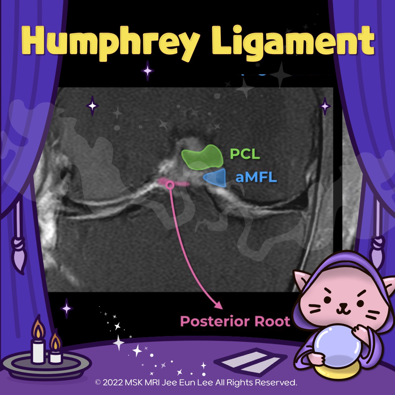
https://youtu.be/jUvkKw5sqvQ https://youtu.be/kEuMzUrHD0M "🧐 Deep Dive into the Meniscofemoral Ligament (MFL) Anatomy! ✨ The MFL, a critical component of the knee, originates from the posterior horn of the lateral meniscus and inserts onto the lateral aspect of the posterior medial femoral condyle. It plays a key role in the stability and function of the knee joint. 🔍 Let's break it down: 1️⃣ Th.. 더보기
[Anatomy_31] Anterior meniscofemoral ligament, ​​Anomalous insertion of the medi Anomalous insertion of the medial meniscus (AIMM) into the anterior cruciate ligament (ACL) is rare.The AIMM has the same anatomical description as the anteromedial meniscofemoral ligament It appears as a linear band isointense to the ACL and meniscus, which runs parallel to the ACL and demonstrates a hyperintense gap between it and the ACL On MRI, this anomaly may be misinterpreted as an ACL te.. 더보기
[Anatomy_30] Oblique Meniscomeniscal Ligament vs bucket handle tear -03 The medial OMML originates from the anterior horn of the medial meniscus and inserts onto the posterior horn of the lateral meniscus between the ACL and PCL. This structure should not be mistaken for a bucket handle meniscal tear A bucket-handle tear is a longitudinal tear with central migration of the inner “handle” fragment The correct diagnosis of an oblique meniscomeniscal ligament rather th.. 더보기
[Anatomy_29] Oblique Meniscomeniscal Ligament -02 The medial OMML originates from the anterior horn of the medial meniscus and extends through the intercondylar notch to insert onto the posterior horn of the lateral meniscus. This ligament passes between the ACL and PCL. This oblique meniscomeniscal ligament is kind of similar SI to meniscal tissue. The sagittal image near the midline demonstrates a linear low signal intensity structure atop th.. 더보기
[Anatomy_28] Oblique Meniscomeniscal Ligament -01 The medial and lateral oblique menisco-meniscal ligaments are rare and can mimic a tear if not recognized. The oblique meniscomeniscal ligament passes from the anterior horn of one meniscus to the posterior horn of the opposite meniscus. The oblique meniscomeniscal ligaments are named based on their anterior attachment site. The medial OMML originates from the anterior horn of the medial meniscu.. 더보기
[Anatomy_27] Anterior transverse meniscal ligament -02 Both horns of the lateral meniscus are triangular in shape and have very sharp points. In this case, we can this low-signal-intensity band like structure adjacent to the anterior horn of the lateral meniscus Both cases are normal finding, The transverse ligament has various diameters. The anterior root of MM inserts along the tibial eminence, and posterior fibers of the anterior horn attachment .. 더보기
[Anatomy_26] Anterior transverse meniscal ligament -01 The transverse ligament is a fibrous band that connects the anterior horns of the medial and lateral menisci. Axial and sagittal images demonstrate the course of the transverse ligament as a low-signal-intensity band traversing Hoffa’s infrapatellar fat pad. The transverse ligament can simulate an oblique tear adjacent to the anterior horn of the lateral meniscus © 2021 MSK MRI Jee Eun Lee All R.. 더보기
[Anatomy_21] Posteroinferior popliteomeniscal fascicle The popliteomeniscal fascicles consist of the anteroinferior(strongest), posterosuperior, and posteroinferior. posteroinferior The posteroinferior popliteomeniscal fascicle (arrow) is the least consistently visualized of the fascicles The posteroinferior fascicle is more medially located, and extends from a medial aponeurotic extension of the popliteus tendon to attach to the lateral meniscus ne.. 더보기