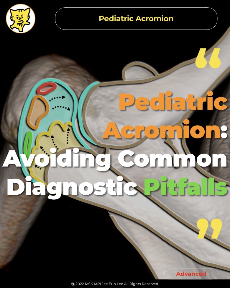
✅ Shoulder MRI 33

Rotator Cable vs. Coracoacromial Ligament on MRI: How to Avoid Common Misinterpretations and Improve Shoulder Tear Diagnosis

https://youtu.be/GLGz8BKbacQ📌 Rotator Cable OverviewThe rotator cable is a thick, fibrous structure in the shoulder, resembling a suspension bridge. It transfers forces across the rotator cuff and helps anchor the tendons to the tubercles of the humerus. ✅ Function of the Rotator CableAnchors rotator cuff tendons to the humerus.Assists in stabilizing the glenohumeral joint.Disperses stress on t..

Shoulder Cysts on MRI: Normal Variant or Hidden Pathology? Bare area cysts

https://youtu.be/XAWfmcwBm9ASummary of Subcortical Cystic Changes of the Humeral Tuberosities  1. IncidenceFound in 15% to 45% of asymptomatic shoulders, often linked to ageing and rotator cuff pathology.  2. ClassificationClassified by location: bare area, lesser tuberosity, anterior greater tuberosity, and posterior greater tuberosity.  3. Bare-Area CystsMost common type, located in the poster..

Supraspinatus Anatomy and Hidden Supraspinatus Myotendinous Injuries

https://youtu.be/jfm5azt1dpA📌  Supraspinatus Anatomy and Hidden Supraspinatus Myotendinous Injuries✅ Supraspinatus Anatomy  The supraspinatus has anterior and posterior components, with the anterior bundle being five times larger.Anterior Bundle: Occupies the anterior supraspinatus fossa and attaches to the anterior superior facet of the greater tuberosity near the rotator cuff interval.Posteri..

Rotator Cuff Insertion: What You Thought You Knew Might Be Wrong!

https://youtu.be/O6UFu8agrkgNormal Supraspinatus and Infraspinatus Insertions1. Overlapping AnatomyThe anterior infraspinatus partially covers the posterolateral supraspinatus.This overlap suggests biomechanical significance in shoulder abduction, especially in cases of isolated supraspinatus tears. 2. Supraspinatus and Infraspinatus FootprintsSupraspinatus: Inserts into the anteromedial area of..

Understanding Infraspinatus Tendon Anatomy_ Key Insights for MRI

https://youtu.be/U2tK1jS6vII Infraspinatus Muscle Anatomy and Tendinous Structure1. Overview of Muscle ArchitectureThe infraspinatus muscle consists of two distinct portions:These portions are connected at the superior muscular area but clearly separate in the distal tendinous region.2. Tendinous CharacteristicsThe tendinous portion is primarily derived from the oblique part:3. Distinctive Attac..

[Advanced] Understanding Rotator Cuff Tendon Layers: Structure and MRI Insights

Five Histologic Layers of the Supraspinatus and Infraspinatus Tendons1. Layer 1: Superficial LayerThickness: ~1 mmComposed of fibers from the coracohumeral ligament.2. Dense Parallel Tendon FibersThickness: 3–5 mmDensely packed parallel collagen fibers.Stronger than layer 3 by twofold.Prone to intrasubstance tears between this layer and layer 3.3. Deep Layer with Non-Uniform Collagen Orientation..

[Advanced] Mastering the Subscapularis Footprint Anatomy

https://youtu.be/pcUqlQzM1LY Summary: Footprint of the Subscapularis1. General Anatomy of the Subscapularis FootprintThe superior footprint of the subscapularis (SSC) tendon is broad and tapered inferiorly.A thin muscular insertion is located below the lesser tuberosity (LT) in the surgical neck of the humerus.The upper two-thirds are subjected to greater tension and mechanical stress.The lower ..

[Advanced] Step 4: Confirm the Presence and Extent of Subscapularis Tears on Sagittal Images

https://youtu.be/5GVpAklu8UE MRI Review: Identifying Subscapularis Tears✅ Understanding Rotator Cuff AnatomySubscapularis Tendon: Attaches to the lesser tuberosity, just anterior to the long head of the biceps tendon (LHB).Greater Tuberosity: Divided into three facets—superior, middle, and inferior. Supraspinatus has a smaller footprint than previously believed; much of this area is occupied by ..

[Essential] Step 3: Evaluating Subscapularis Tendon Tears with Muscle Atrophy on Sagittal Images

https://youtu.be/Ilza4mNbkxgSubscapularis Muscle Anatomy and FunctionThe subscapularis is innervated by both the upper and lower subscapular nerves.Primary actions: internal rotation and adduction of the shoulder.Fatty Infiltration as an IndicatorFatty infiltration of the subscapularis muscle is a specific but insensitive marker for subscapularis tendon tears.The variability of this finding rela..

[Essential] ​​Step 2 - Subscapularis Tears and Biceps Tendon Medial Dislocation

https://youtu.be/aUJeM0sB8aU✅ Indirect Imaging Findings of Subscapularis Tendon Tear1️⃣ Medial Displacement of Long Head Biceps TendonMedial dislocations or subluxations of the long head biceps brachii tendon are commonly associated with subscapularis tendon tears.Intraarticular dislocation of the long head biceps tendon suggests a full-thickness tear of the tendinous attachment to the lesser tu..