✨ Join the channel to enjoy the benefits! 🚀https://www.youtube.com/channel/UC4bw7o0l2rhxn1GJZGDmT9w/join
https://youtube.com/shorts/tSPHy2Amd8I
- YouTube
www.youtube.com
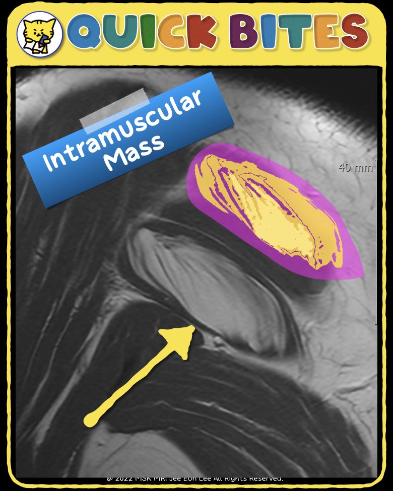
📌Intramuscular Lipoma: Key Imaging Features
Definition
Intramuscular lipomas are deep-seated benign lipomatous tumors located within a muscle.
✅CT Features
- Hypodense Soft Tissue Mass: Appears as a low-density mass within the musculature.
- Fat Density: Typically shows Hounsfield Unit (HU) measurements in the negative range.
- Striated Appearance: Some cases exhibit a striated texture.
- Intramuscular Septae & Interdigitations: May present with thick septae and an interdigitating pattern.
- Shape Variability: Commonly oval or fusiform but may vary.
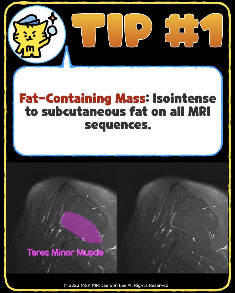
✅MRI Features
- Fat-Containing Mass: Isointense to subcutaneous fat in all sequences.
- Septae Presence: Thin septae may be observed but should not enhance avidly or show nodules.
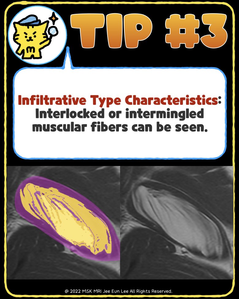
- Infiltrative Type Characteristics: Interlocked or intermingled muscular fibers can be seen.
- Capsule Consideration: If a capsule is present outside the mass, muscular fibers should not be part of the main lesion.
#IntramuscularLipoma, #MSKRadiology, #FatDensity, #Lipoma, #SoftTissueTumor,
"Visualizing MSK Radiology: A Practical Guide to Radiology Mastery"
© 2022 MSK MRI Jee Eun Lee All Rights Reserved.
No unauthorized reproduction, redistribution, or use for AI training.