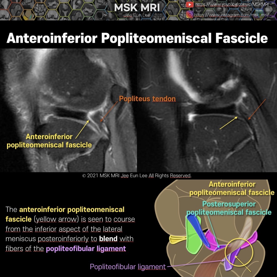
The anteroinferior popliteomeniscal fascicle (yellow arrow) is seen to course from the inferior aspect of the lateral meniscus posteroinferiorly to blend with fibers of the popliteofibular ligament
This fascicle is typically seen on sagittal slices where the fibular head is also visible.
In another patient, a component of the anteroinferior popliteomeniscal fascicle which courses directly to the popliteus tendon.
On this sagittal image, the posterosuperior fascicle is not seen normally. Because the posterosuperior fascicle is located more medially to the anteroinferior fascicle.
This component is usually inconsistently visible on routine sagittal MR images
#MSKMRI, #virtualMRI, #radiologist, #Knee_MRI, #MSKMRI_Knee, #Knee_anatomy, #Knee_meniscus, #meniscus, #Virtual_MRI, #MRI_illustrator, #lateralmeniscus, #LM, #popliteomeniscalfascicle, #lateralmeniscustear,
#popliteo #popliteomeniscalfascicle #posterolateralcorner
It's not a real patient's MRI, but they are virtual images very similar to the images in the journals. The images will be created for educational purposes.
All copyrights belong to MSK MRI Jee Eun Lee.
You may not distribute or commercially exploit the content. Nor may you transmit it or store it on any other website or other forms of the electronic retrieval system.
If you would like to use an image or video for anything other than personal use, please contact me. (jamaisvu1977@gmail.com)
#Virtual MRI, #MRI illustrator, #MSKMRI © 2021 MSK MRI Jee Eun Lee All Rights Reserved.

'Knee MRI > Meniscus' 카테고리의 다른 글
| [Anatomy_20] Posterosuperior popliteomeniscal fascicle (0) | 2021.09.25 |
|---|---|
| [Anatomy_19] Anteroinferior popliteomeniscal fascicle 02 (0) | 2021.09.25 |
| [Anatomy_17] Tear of posterosuperior Popliteomeniscal fascicle (0) | 2021.09.25 |
| [Shorts #02] Popliteomeniscal fascicle, #shorts (0) | 2021.09.24 |
| [Anatomy_16] Overview of the popliteomeniscal fascicles (0) | 2021.09.24 |