The anteroinferior and posterosuperior fascicles were seen on MRI in 97% of patients who had a normal lateral meniscus at arthroscopy
Although 45 degree oblique coronal images have been recommended for visualization of the popliteomeniscal fascicles , these structures are often well visualized during the review of routine sagittal acquisitions of the knee
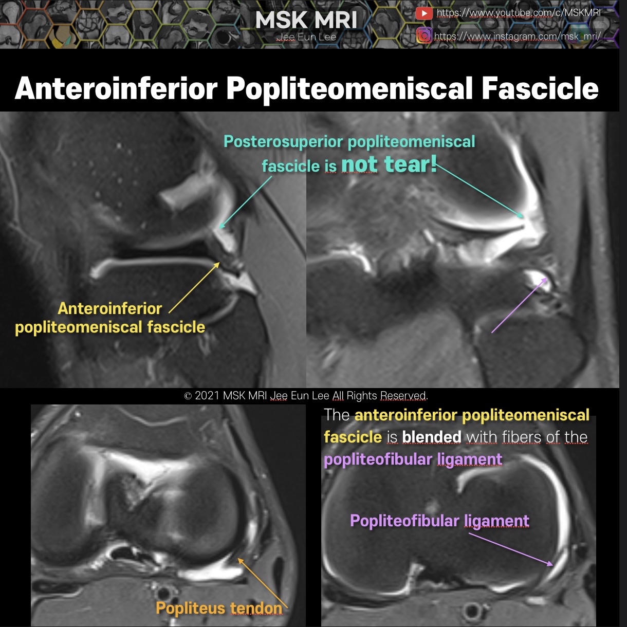
The anteroinferior popliteomeniscal fascicle is seen to course from the inferior aspect of the lateral meniscus posteroinferiorly to blend with fibers of the popliteofibular ligament
The posterosuperior fascicle is not seen normally. Because the posterosuperior fascicle is located more medially to the anteroinferior fascicle.
It's not a real patient's MRI, but they are virtual images very similar to the images in the journals. The images will be created for educational purposes.
All copyrights belong to MSK MRI Jee Eun Lee.
You may not distribute or commercially exploit the content. Nor may you transmit it or store it on any other website or other forms of the electronic retrieval system.
If you would like to use an image or video for anything other than personal use, please contact me. (jamaisvu1977@gmail.com)
#Virtual MRI, #MRI illustrator, #MSKMRI © 2021 MSK MRI Jee Eun Lee All Rights Reserved.

#MSKMRI, #virtualMRI, #radiologist, #Knee_MRI, #MSKMRI_Knee, #Knee_anatomy, #Knee_meniscus, #meniscus, #Virtual_MRI, #MRI_illustrator, #lateralmeniscus, #LM, #popliteomeniscalfascicle, #lateralmeniscustear,
#popliteo #popliteomeniscalfascicle #posterolateralcorner
'Knee MRI > Meniscus' 카테고리의 다른 글
| [Anatomy_21] Posteroinferior popliteomeniscal fascicle (0) | 2021.09.25 |
|---|---|
| [Anatomy_20] Posterosuperior popliteomeniscal fascicle (0) | 2021.09.25 |
| [Anatomy_18] Anteroinferior popliteomeniscal fascicle 01 (0) | 2021.09.25 |
| [Anatomy_17] Tear of posterosuperior Popliteomeniscal fascicle (0) | 2021.09.25 |
| [Shorts #02] Popliteomeniscal fascicle, #shorts (0) | 2021.09.24 |