The popliteomeniscal fascicles consist of the anteroinferior(strongest), posterosuperior, and posteroinferior.
posteroinferior
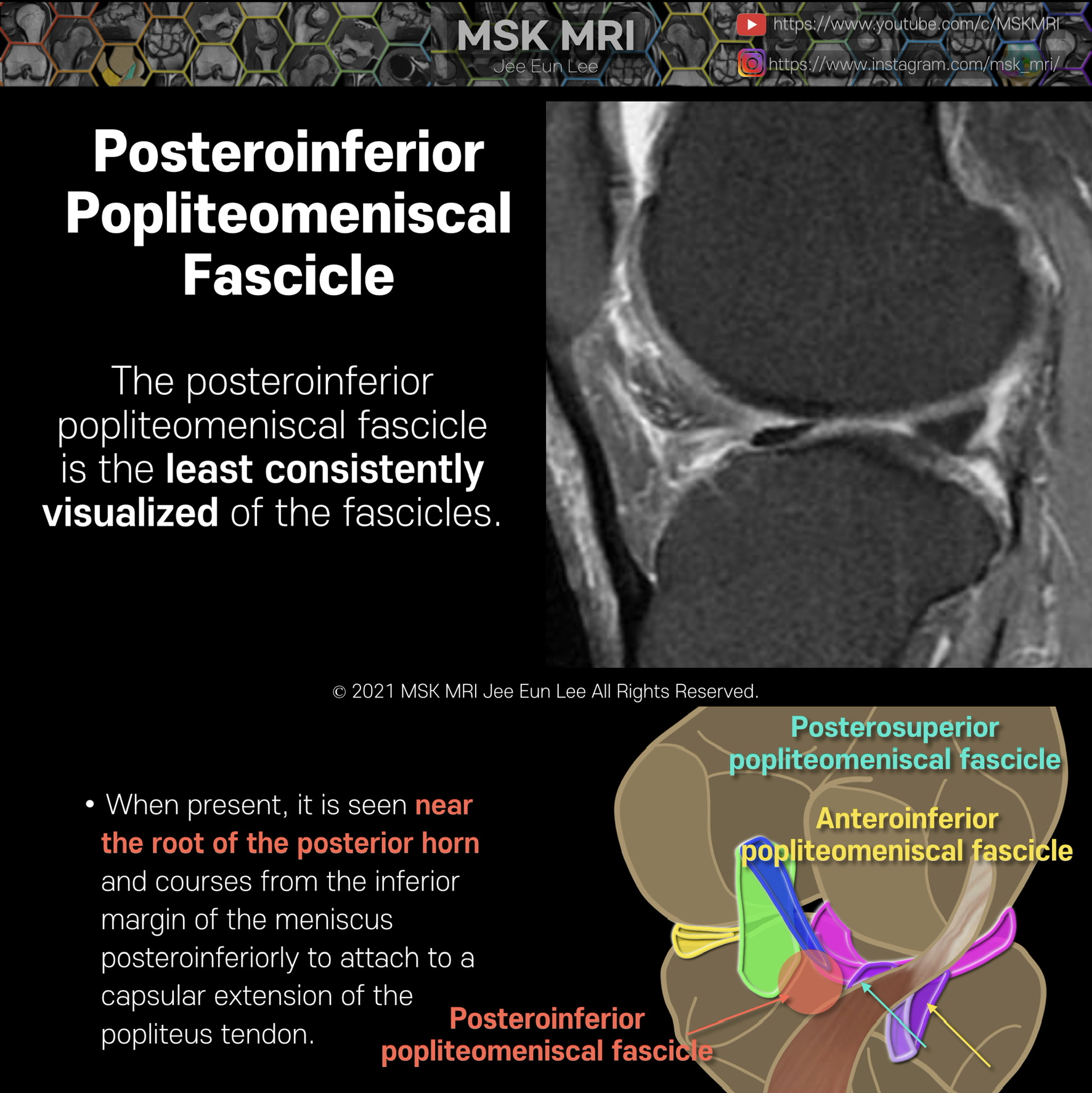
The posteroinferior popliteomeniscal fascicle (arrow) is the least consistently visualized of the fascicles
The posteroinferior fascicle is more medially located, and extends from a medial aponeurotic extension of the popliteus tendon to attach to the lateral meniscus near the attachment of the meniscofemoral ligaments.
it may resemble an inferior torn flap of the meniscus
© 2021 MSK MRI Jee Eun Lee All Rights Reserved.
#MSKMRI, #virtualMRI, #radiologist, #Knee_MRI, #MSKMRI_Knee, #Knee_anatomy, #Knee_meniscus, #meniscus, #Virtual_MRI, #MRI_illustrator, #lateralmeniscus, #LM, #popliteomeniscalfascicle, #lateralmeniscustear,


'Knee MRI > Meniscus' 카테고리의 다른 글
| [Anatomy_22] Meniscofemoral ligament, Humphry, Wrisberg (0) | 2021.09.26 |
|---|---|
| [Shorts #03] Meniscofemoral ligament, Humphry, Wrisberg #shorts (0) | 2021.09.25 |
| [Anatomy_20] Posterosuperior popliteomeniscal fascicle (0) | 2021.09.25 |
| [Anatomy_19] Anteroinferior popliteomeniscal fascicle 02 (0) | 2021.09.25 |
| [Anatomy_18] Anteroinferior popliteomeniscal fascicle 01 (0) | 2021.09.25 |