==============================================
⬇️✨⬇️🎉⬇️🔥⬇️📚⬇️
Click the link to purchase on Amazon 🎉📚
==============================================
🎥 Check Out All Videos at Once! 📺
👉 Visit Visualizing MSK Blog to explore a wide range of videos! 🩻
https://visualizingmsk.blogspot.com/?view=magazine
📚 You can also find them on MSK MRI Blog and Naver Blog! 📖
https://www.instagram.com/msk_mri/
Click now to stay updated with the latest content! 🔍✨
==============================================
✅ Hypermobile Lateral Meniscus:
- It is thought to result from the congenital absence of posterior capsular attachments (similar to Wrisberg-type discoid meniscus, but without discoid morphology) or from tears in the posterior capsular attachment, especially the popliteomeniscal fascicles.
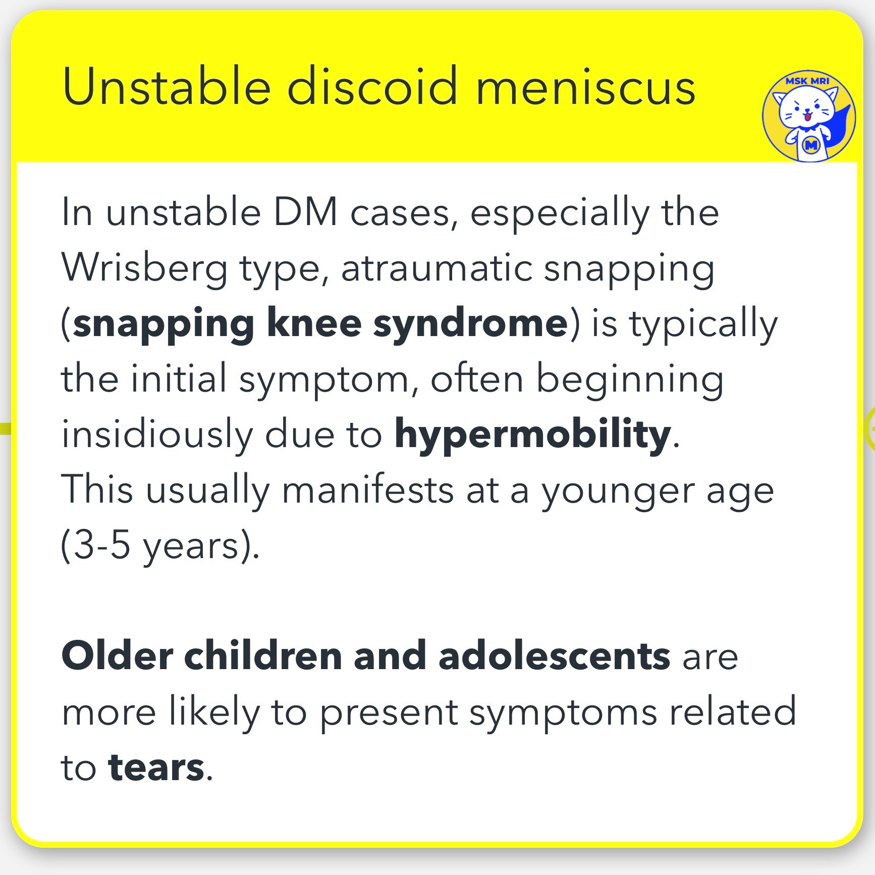
✅ Unstable Discoid Meniscus (DM):
- Symptoms: In unstable DM cases, including the Wrisberg type, symptoms typically include atraumatic snapping of insidious onset (snapping knee syndrome), presenting at a younger age (3–5 years). Older children and adolescents often present with tear-related symptoms.
✅ Early Signs of Instability:
- Indicators: Shape deformation in the discoid meniscus is an early sign of instability, which may include surface changes, meniscal shifts, and meniscal megahorns.
- Imaging Signs: "Pseudo–bucket-handle tear," "crimped meniscus sign," and parameniscal edema are key imaging signs of meniscal instability.
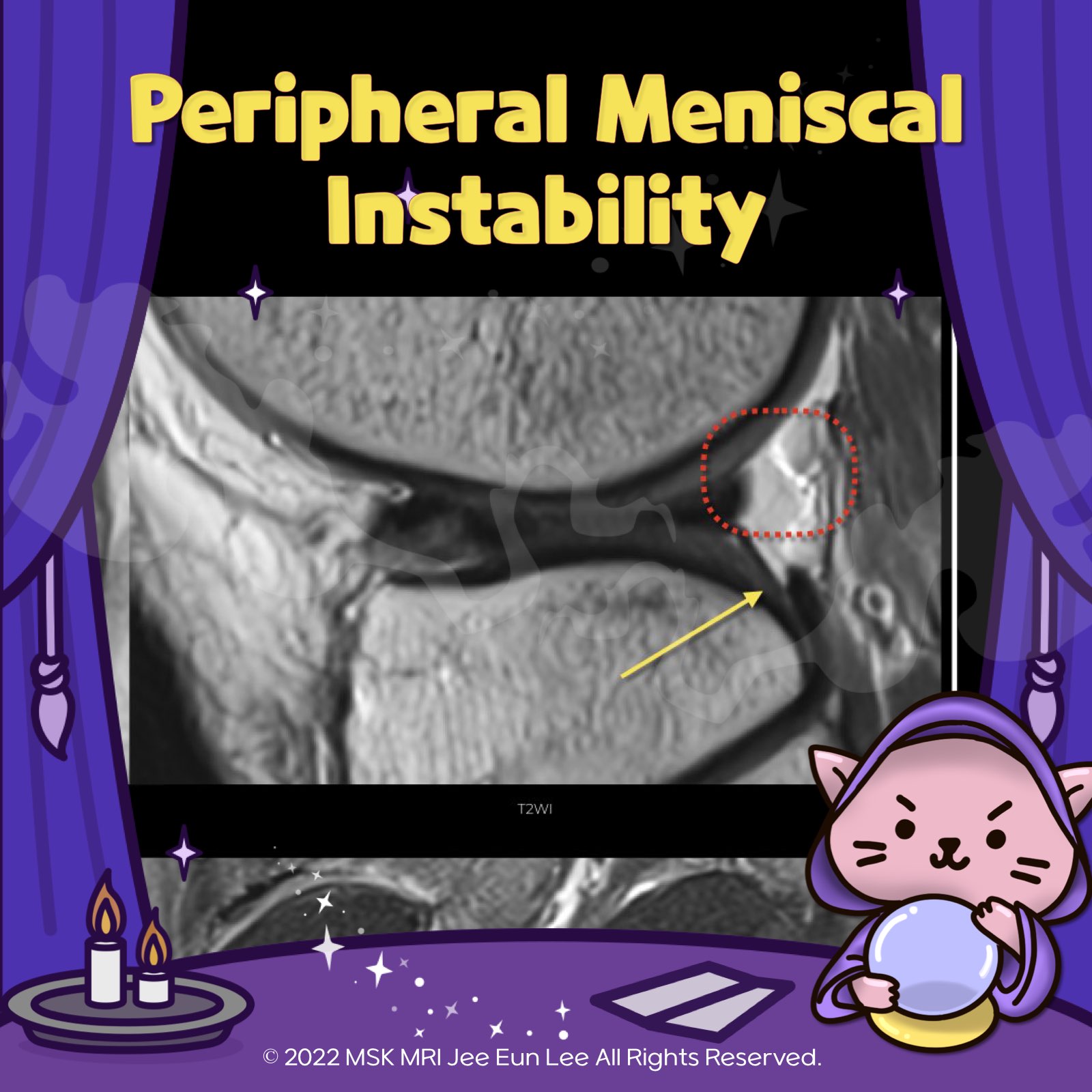
✅ MRI Signs of Peripheral Instability:
- Key Signs: MRI indicators include the absence of capsular insertions (observable as an absence of normal fascicles; T2 signal increase due to lack of coronal ligaments, simulating peripheral rupture) and anterior displacement of the posterior horn of the meniscus relative to the tibia (indicating meniscus subluxation).
"Visualizing MSK Radiology: A Practical Guide to Radiology Mastery"
© 2022 MSK MRI Jee Eun Lee All Rights Reserved.
#VisualizingMSK #Lateralmeniscus #Discoidmeniscus #Meniscaltears #Poplitealmeniscacal #Meniscalinstability


'✅ Knee MRI Mastery > Chap 1. Meniscus' 카테고리의 다른 글
| (Fig 1-C.12) Meniscal Cysts of lateral meniscus (0) | 2024.02.08 |
|---|---|
| (Fig 1-C.11) Meniscal Cysts of medial meniscus (0) | 2024.02.08 |
| (Fig 1-C.08) Degenerated and torn lateral discoid meniscus (0) | 2024.02.08 |
| (Fig 1-C.07) Meniscal shape deformation and torn lateral discoid meniscus (1) | 2024.02.08 |
| (Fig 1-C.06) Ring-shaped meniscus versus bucket handle tear (0) | 2024.02.08 |