Click the link to purchase on Amazon 🎉📚
==============================================
🎥 Check Out All Videos at Once! 📺
👉 Visit Visualizing MSK Blog to explore a wide range of videos! 🩻
https://visualizingmsk.blogspot.com/?view=magazine
📚 You can also find them on MSK MRI Blog and Naver Blog! 📖
https://www.instagram.com/msk_mri/
Click now to stay updated with the latest content! 🔍✨
==============================================
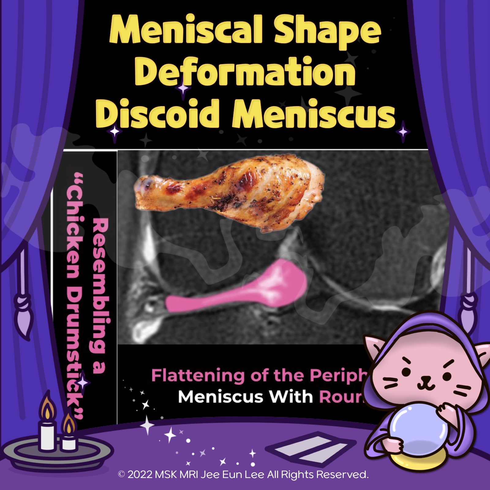
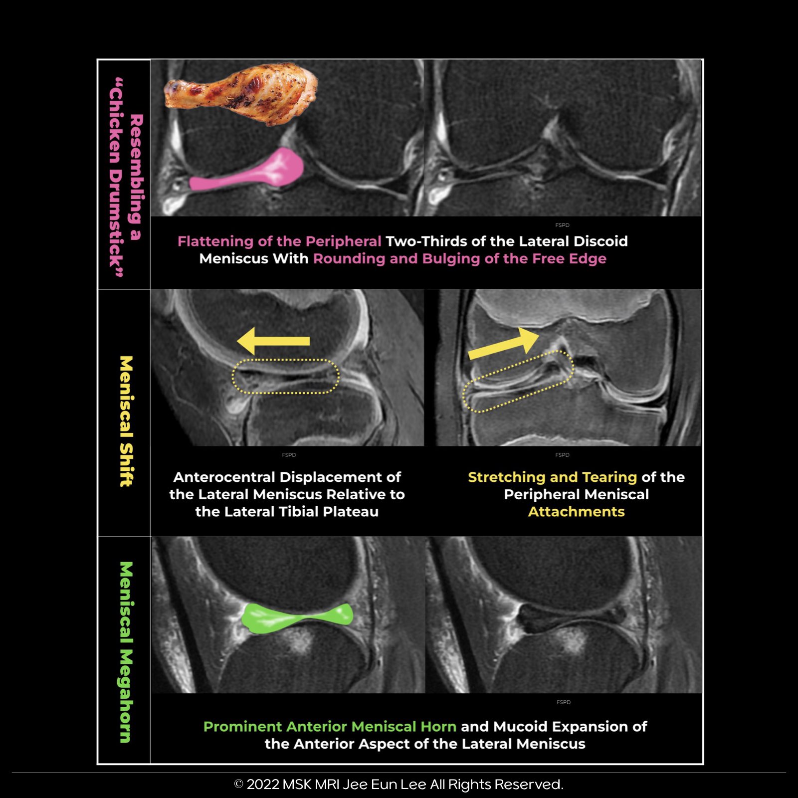
Meniscal and Shape Deformation in Discoid Lateral Meniscus:
- Meniscal Deformation: Indicates peripheral detachment through abnormal infolding, buckling, and shifting.
- Shape Deformation: Signals early instability with surface changes, meniscal shifts, and development of meniscal megahorns. Important imaging signs include:
Additional Key Features:
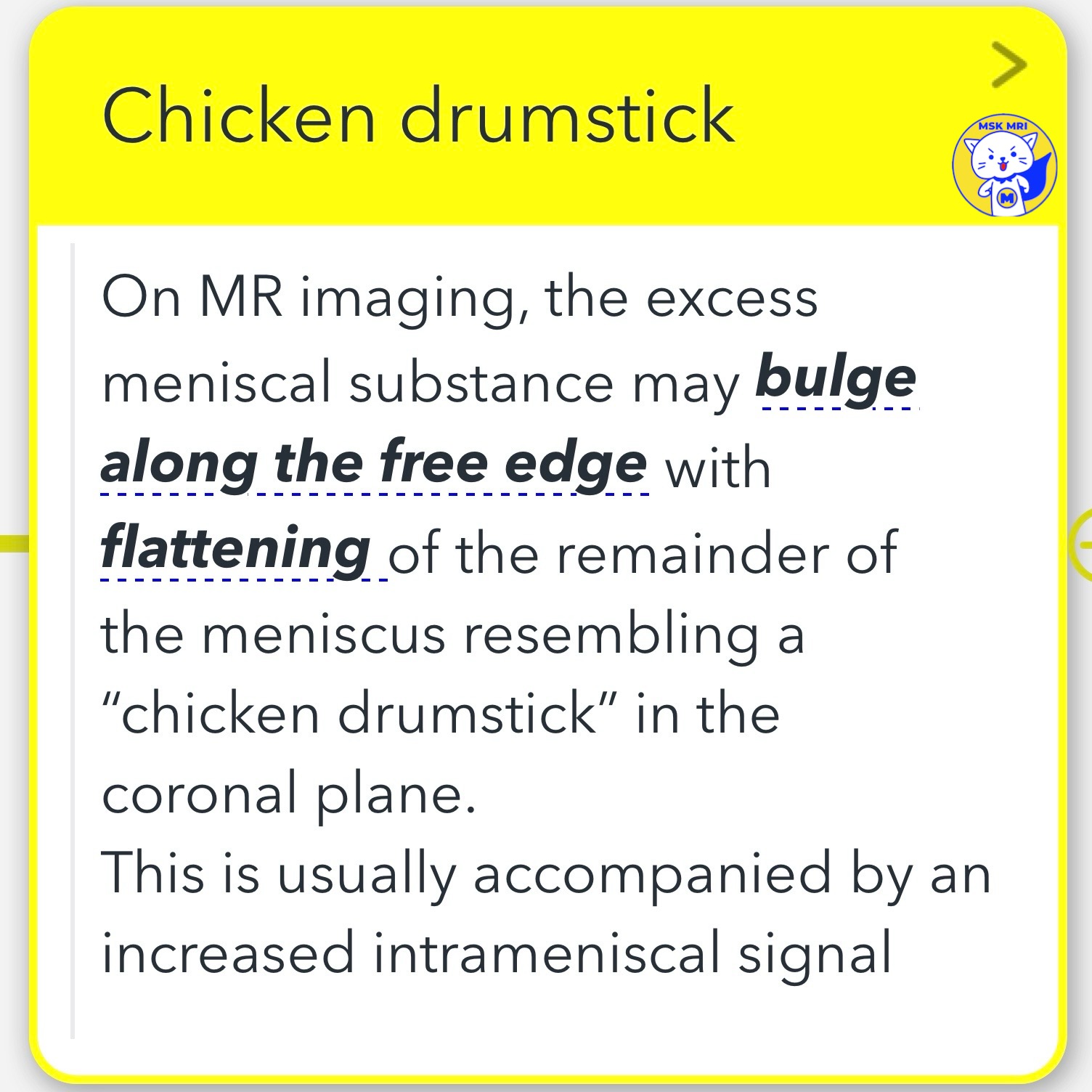
- Chicken Drumstick Appearance:
On MRI, the meniscus may bulge at the free edge and flatten elsewhere, resembling a chicken drumstick in the coronal plane, often with an increased intrameniscal signal. - Pseudo–Bucket-Handle Tear:
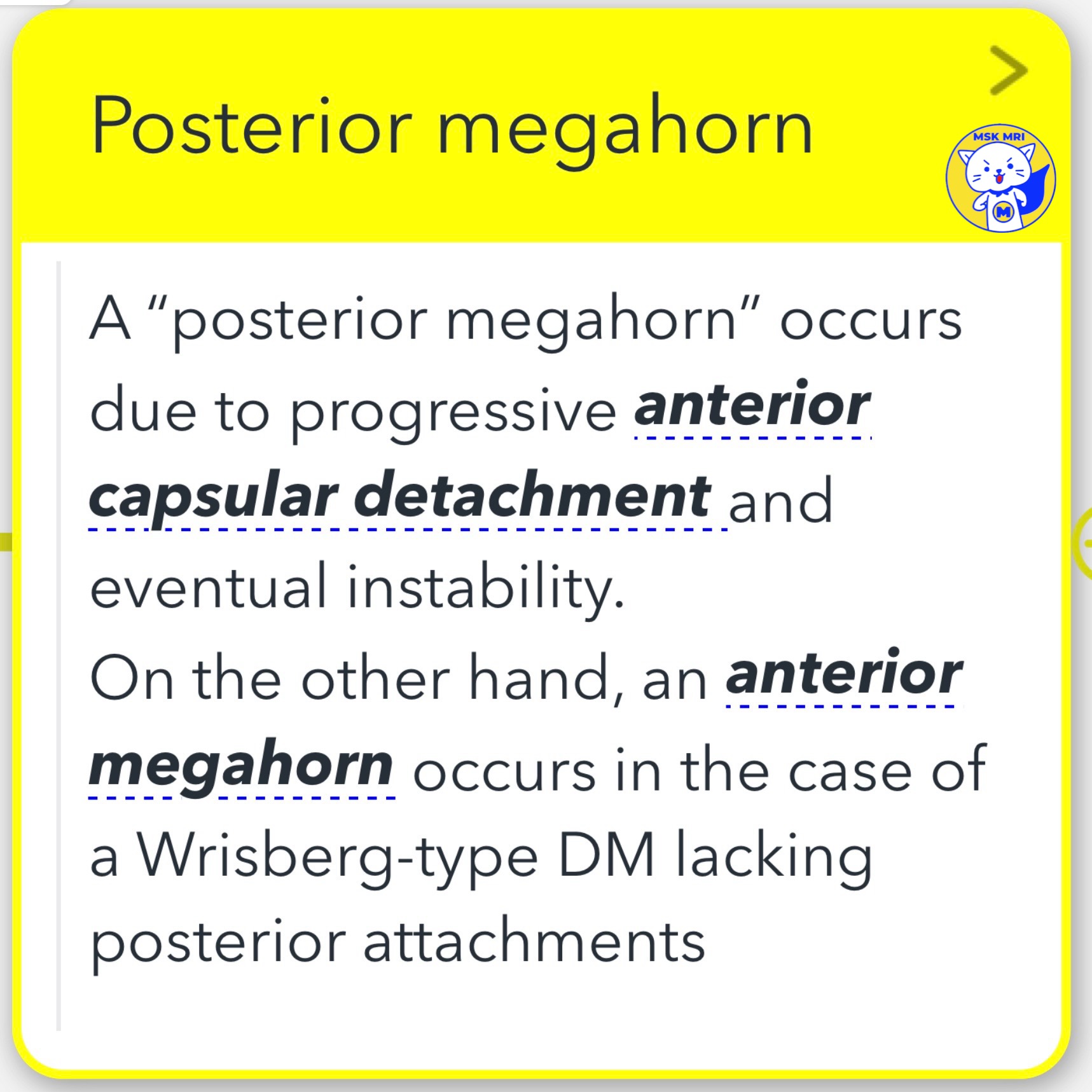
Refers to a centrally flipped discoid meniscus (DM) or middle segment into the intercondylar recess without a longitudinal tear, indicating underlying hypermobility and peripheral rim instability. - Posterior Megahorn:
Results from progressive anterior capsular detachment leading to instability. Conversely, an anterior megahorn indicates a Wrisberg-type DM with absent posterior attachments.
Restrepo R, Weisberg MD, Pevsner R, Swirsky S, Lee EY. Discoid Meniscus in the Pediatric Population:: Emphasis on MR Imaging Signs of Instability. Magn Reson Imaging Clin N Am. 2019 May;27(2):323-339
"Visualizing MSK Radiology: A Practical Guide to Radiology Mastery"
© 2022 MSK MRI Jee Eun Lee All Rights Reserved.
#VisualizingMSK #Lateralmeniscus #Discoidmeniscus #Meniscaltears #Wrisbergmeniscus #Meniscalinstability


'✅ Knee MRI Mastery > Chap 1. Meniscus' 카테고리의 다른 글
| (Fig 1-C.10) Peripheral Meniscal Instability (0) | 2024.02.08 |
|---|---|
| (Fig 1-C.08) Degenerated and torn lateral discoid meniscus (0) | 2024.02.08 |
| (Fig 1-C.06) Ring-shaped meniscus versus bucket handle tear (0) | 2024.02.08 |
| (Fig 1-C.05) Type III Wrisberg-type discoid meniscus (0) | 2024.02.08 |
| (Fig 1-C.04) Type II Incomplete discoid meniscus (0) | 2024.02.08 |