==============================================
⬇️✨⬇️🎉⬇️🔥⬇️📚⬇️
Click the link to purchase on Amazon 🎉📚
==============================================
🎥 Check Out All Videos at Once! 📺
👉 Visit Visualizing MSK Blog to explore a wide range of videos! 🩻
https://visualizingmsk.blogspot.com/?view=magazine
📚 You can also find them on MSK MRI Blog and Naver Blog! 📖
https://www.instagram.com/msk_mri/
Click now to stay updated with the latest content! 🔍✨
==============================================
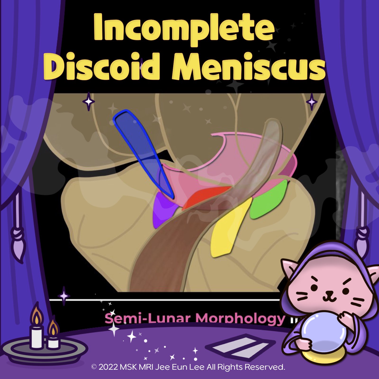
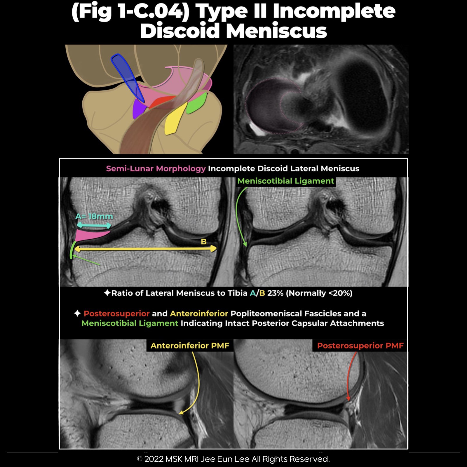
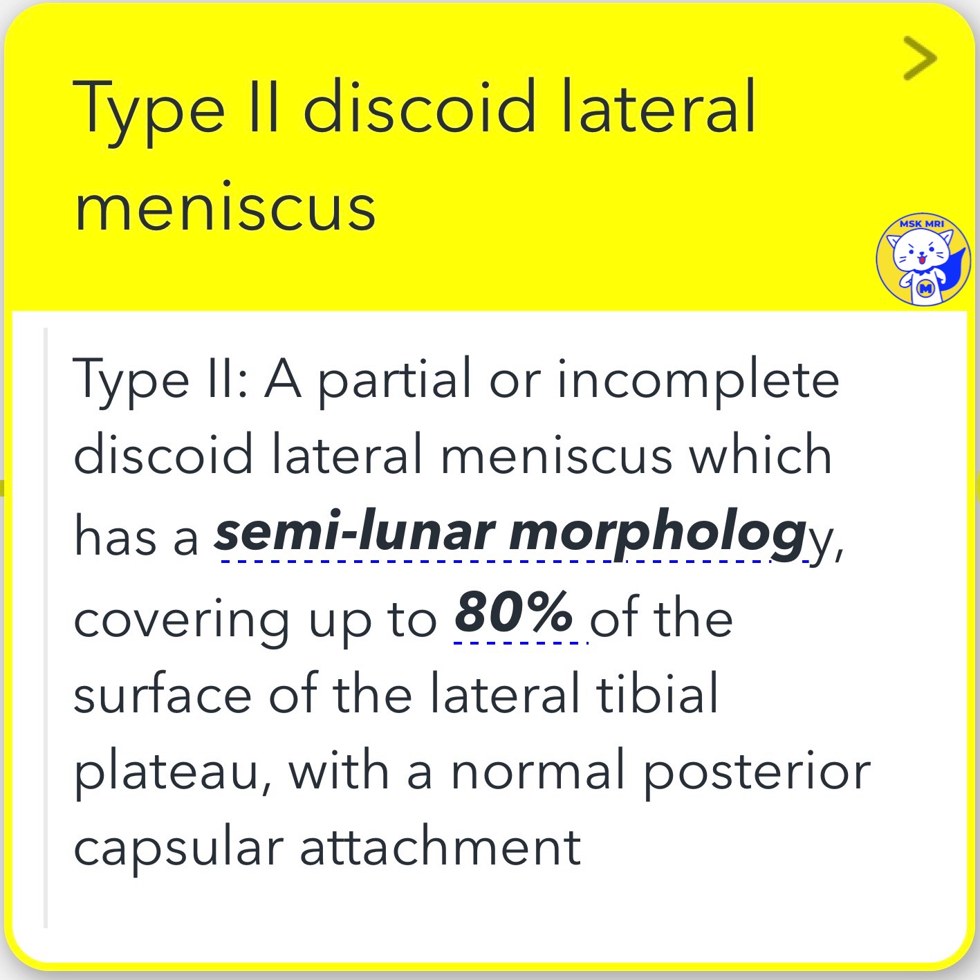
Type II: partial or incomplete discoid lateral meniscus
A partial or incomplete discoid lateral meniscus that has a semi-lunar morphology, covering up to 80% of the surface of the lateral tibial plateau, with a normal posterior capsular attachment
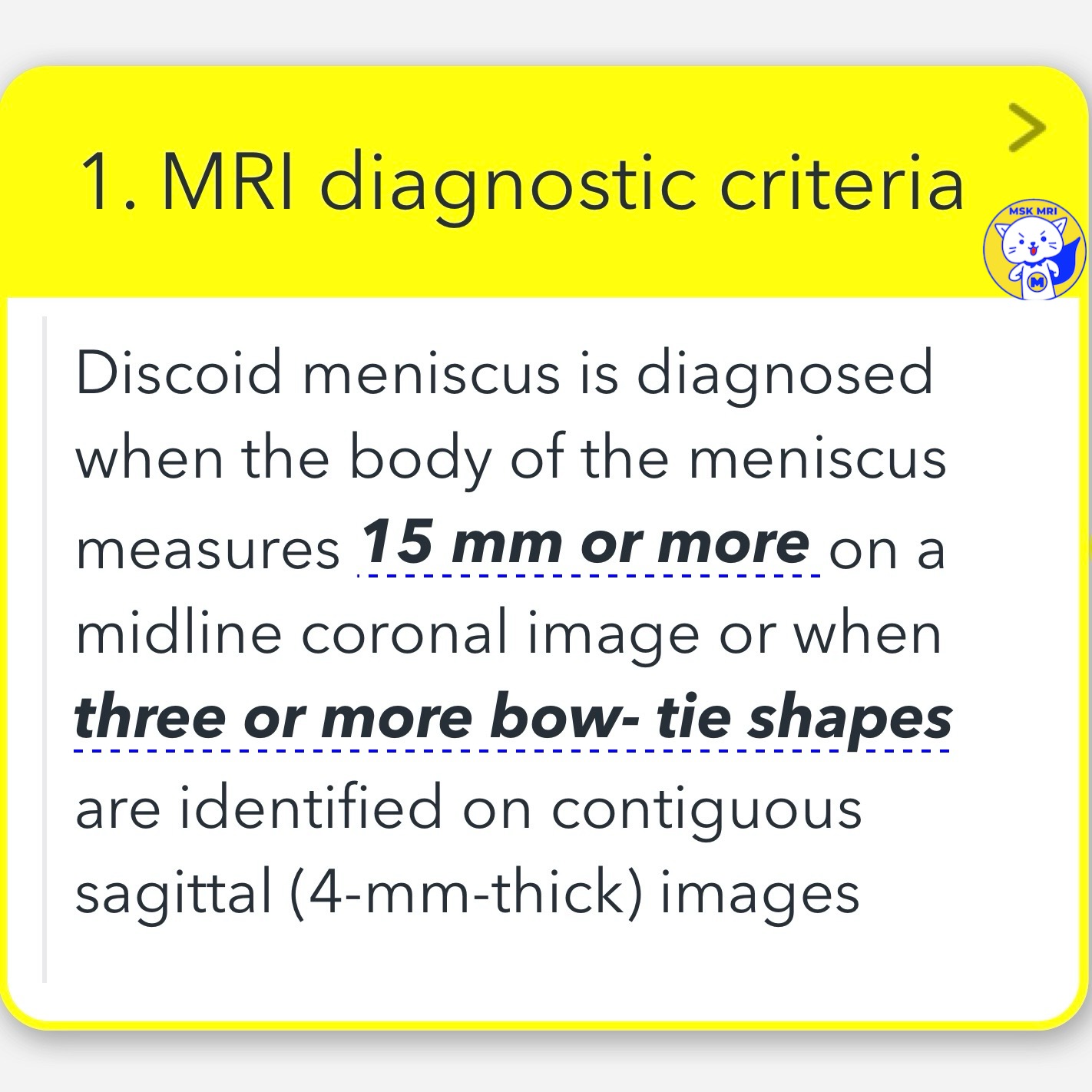
1. MRI diagnostic criteria of Discoid meniscus
- Discoid meniscus is diagnosed when the body of the meniscus measures 15 mm or more on a midline coronal image or when three or more bow-tie shapes are identified on contiguous sagittal (4-mm-thick) images
2. MRI diagnostic criteria of Discoid meniscus
- The ratio of lateral meniscus-to-tibia (RMT) width is a more accurate method to determine the presence of a discoid lateral meniscus on MRI.
- The ratio of the minimal meniscal width to the maximal tibial width of more than 20% may be used.
"Visualizing MSK Radiology: A Practical Guide to Radiology Mastery"
© 2022 MSK MRI Jee Eun Lee All Rights Reserved.
#VisualizingMSK #Lateralmeniscus #Discoidmeniscus #Meniscaltears #IncompleteDiscoid

'✅ Knee MRI Mastery > Chap 1. Meniscus' 카테고리의 다른 글
| (Fig 1-C.06) Ring-shaped meniscus versus bucket handle tear (0) | 2024.02.08 |
|---|---|
| (Fig 1-C.05) Type III Wrisberg-type discoid meniscus (0) | 2024.02.08 |
| (Fig 1-C.03) Type I Complete discoid meniscus (1) | 2024.02.08 |
| (Fig 1-B.51) Medial and Lateral Meniscocapsular Separations (0) | 2024.02.08 |
| (Fig 1-B.50) Medial Meniscocapsular Separation (0) | 2024.02.07 |