Click the link to purchase on Amazon 🎉📚
==============================================
🎥 Check Out All Videos at Once! 📺
👉 Visit Visualizing MSK Blog to explore a wide range of videos! 🩻
https://visualizingmsk.blogspot.com/?view=magazine
📚 You can also find them on MSK MRI Blog and Naver Blog! 📖
https://www.instagram.com/msk_mri/
Click now to stay updated with the latest content! 🔍✨
==============================================
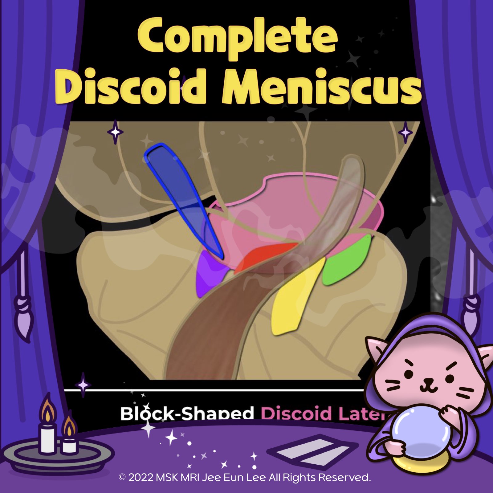
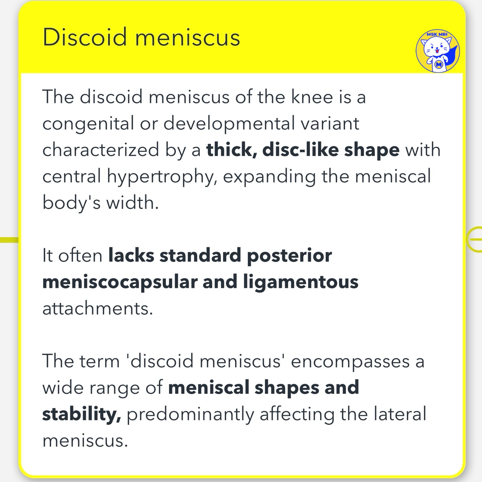
🤔Discoid Meniscus Overview:
- The discoid meniscus of the knee is a congenital or developmental variant with a thick, disc-like shape and central hypertrophy, leading to an increased width of the meniscal body.
- It typically lacks standard posterior meniscocapsular and ligamentous attachments, with the ‘discoid meniscus’ term covering a broad spectrum of shapes and stability levels, mainly affecting the lateral meniscus.
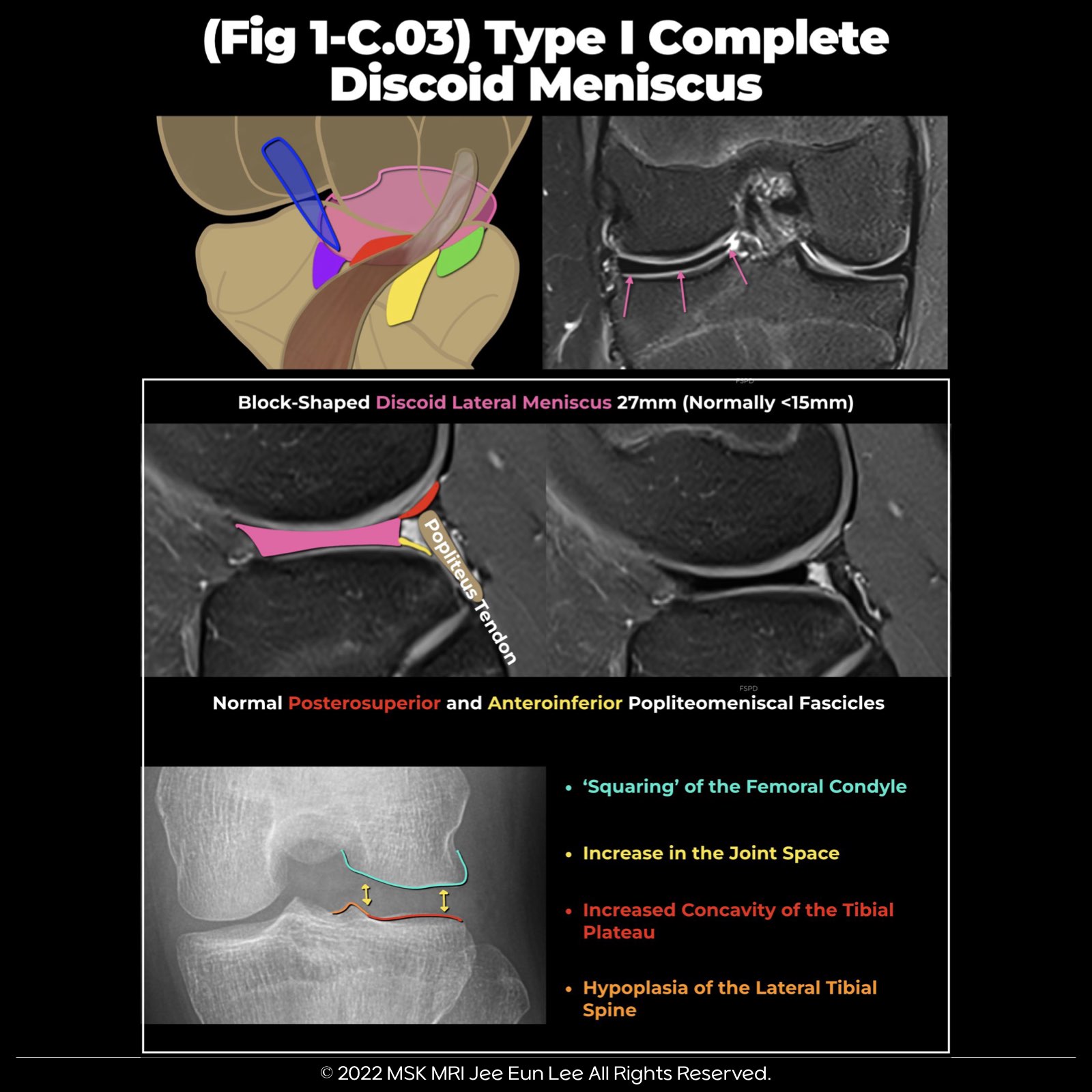
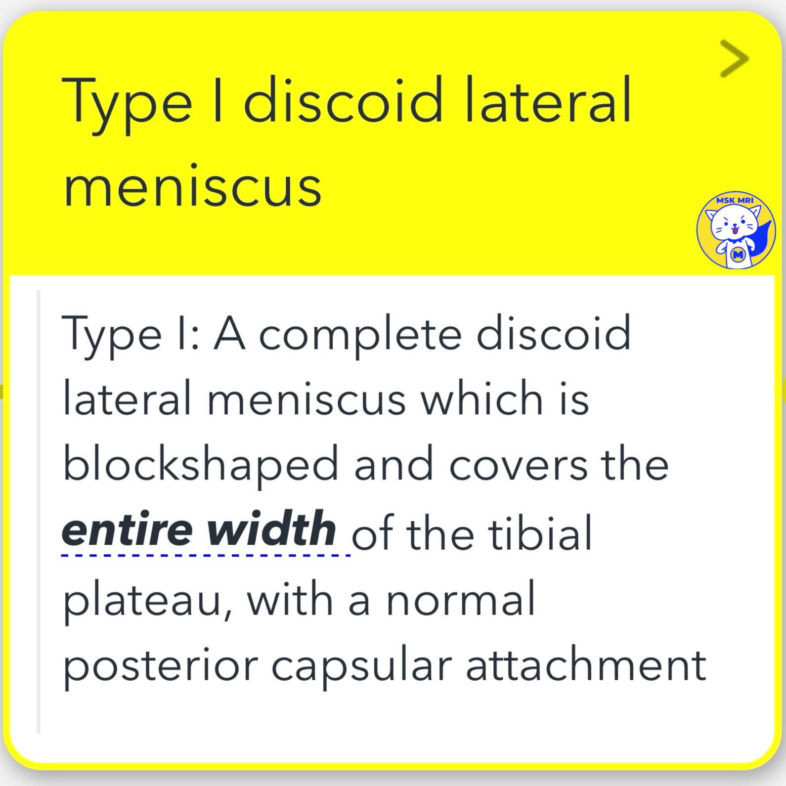
🤔Type I:
- A complete discoid lateral meniscus, characterized by a block-shaped structure that spans the entire tibial plateau width, maintaining normal posterior capsular attachment.
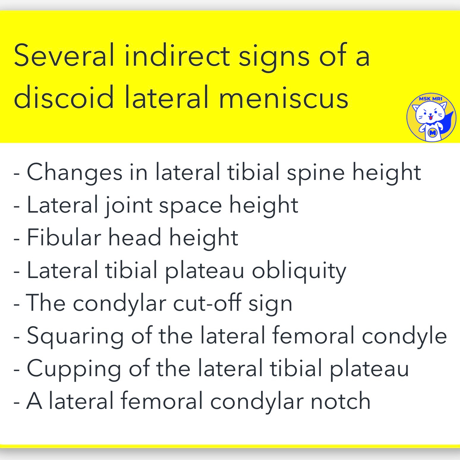
🤔 Several indirect signs of a discoid lateral meniscus
- Changes in lateral tibial spine height
- Lateral joint space height
- Fibular head height
- Lateral tibial plateau obliquity
- The condylar cut-off sign
- Squaring of the lateral femoral condyle
- Cupping of the lateral tibial plateau
- A lateral femoral condylar notch
"Visualizing MSK Radiology: A Practical Guide to Radiology Mastery"
© 2022 MSK MRI Jee Eun Lee All Rights Reserved.
#VisualizingMSK #Lateralmeniscus #Discoidmeniscus #Meniscaltears #CompleteDiscoid
'✅ Knee MRI Mastery > Chap 1. Meniscus' 카테고리의 다른 글
| (Fig 1-C.05) Type III Wrisberg-type discoid meniscus (0) | 2024.02.08 |
|---|---|
| (Fig 1-C.04) Type II Incomplete discoid meniscus (0) | 2024.02.08 |
| (Fig 1-B.51) Medial and Lateral Meniscocapsular Separations (0) | 2024.02.08 |
| (Fig 1-B.50) Medial Meniscocapsular Separation (0) | 2024.02.07 |
| (Fig 1-E.49) Isolated Tear of popliteomeniscal fascicles (0) | 2024.02.07 |