==============================================
⬇️✨⬇️🎉⬇️🔥⬇️📚⬇️
Click the link to purchase on Amazon 🎉📚
==============================================
🎥 Check Out All Videos at Once! 📺
👉 Visit Visualizing MSK Blog to explore a wide range of videos! 🩻
https://visualizingmsk.blogspot.com/?view=magazine
📚 You can also find them on MSK MRI Blog and Naver Blog! 📖
https://www.instagram.com/msk_mri/
Click now to stay updated with the latest content! 🔍✨
==============================================
🔵🇰🇷Tear of popliteomeniscal fascicles
- When assessing a knee for a hypermobile lateral meniscus (LM), it's important for the radiologist to concentrate on the anteroinferior and posterosuperior popliteomeniscal fascicles (PMFs).
- This is because a total rupture or lack of these structures correlates with a significantly higher likelihood (with odds ratios of 12 and 6, respectively) of a hypermobile LM.
- Despite some controversy, there are surgeons who advocate for repairing all instances of symptomatic LM instability when an MRI and arthroscopy confirm a PMF tear.
Radiographics. 2023 Jul;43(7):e220208.
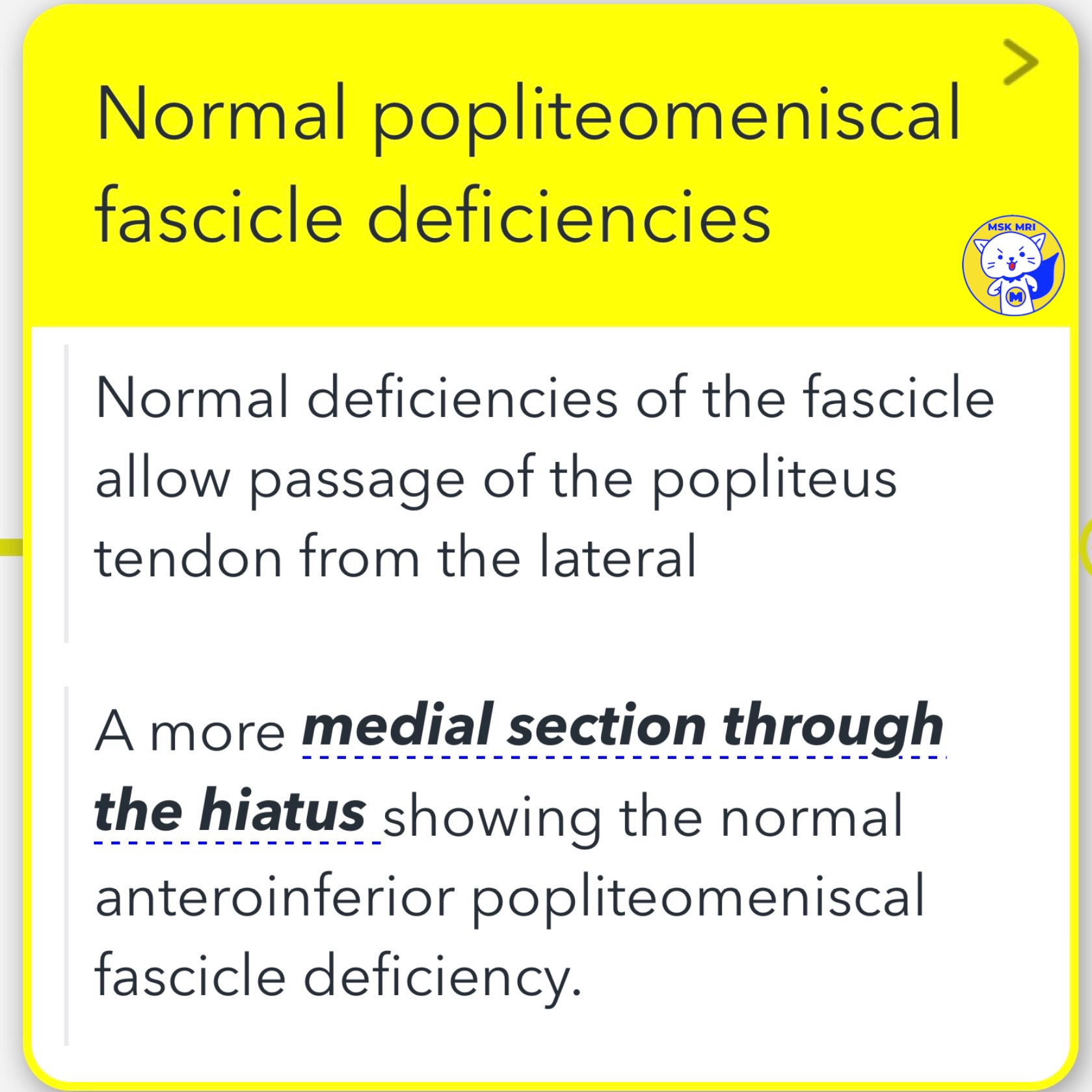
🔵🇰🇷Normal popliteomeniscal fascicle deficiencies
- The normal absence of the anteroinferior popliteomeniscal fascicle allows the popliteus tendon to traverse from the lateral to the medial side through the hiatus.
- A slice further medially through the hiatus reveals this normal deficiency of the fascicle.
"Visualizing MSK Radiology: A Practical Guide to Radiology Mastery"
© 2022 MSK MRI Jee Eun Lee All Rights Reserved.
#VisualizingMSK #Meniscocapsularseparation #PopliteomeniscalFascicles #Lateralmeniscus