==============================================
⬇️✨⬇️🎉⬇️🔥⬇️📚⬇️
Click the link to purchase on Amazon 🎉📚
==============================================
🎥 Check Out All Videos at Once! 📺
👉 Visit Visualizing MSK Blog to explore a wide range of videos! 🩻
https://visualizingmsk.blogspot.com/?view=magazine
📚 You can also find them on MSK MRI Blog and Naver Blog! 📖
https://www.instagram.com/msk_mri/
Click now to stay updated with the latest content! 🔍✨
==============================================
🔴🇰🇷Summary of the information regarding lateral meniscocapsular separation:
- 1️⃣Occurrence with Fractures and ACL Tear:
- Lateral meniscocapsular separation can occur alone or in conjunction with tibia or femur fractures or an ACL tear. The focus here is on cases where it occurs with lateral femoral condyle fractures.
- 2️⃣Injuries in Tibial Plateau Fractures and ACL Ruptures:
- There is a known occurrence of injuries at the lateral meniscocapsular junction region in patients with tibial plateau fractures and ACL ruptures.
- 3️⃣MRI Indicators: In cases of tibial plateau fracture, MRI may suggest lateral meniscocapsular separation when there is:
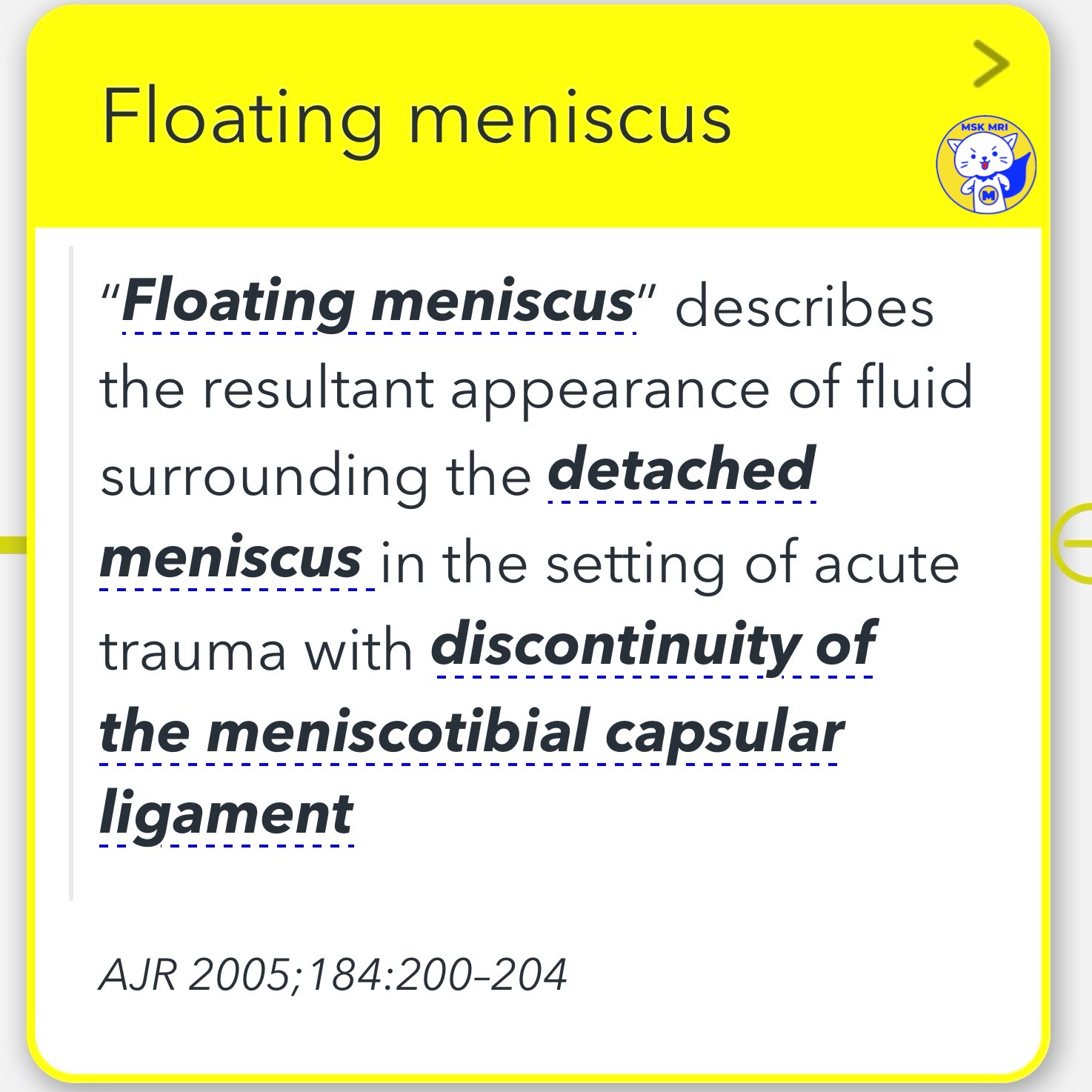
- 4️⃣"Floating Meniscus" Appearance:
- This term describes the appearance of fluid surrounding a detached meniscus in acute trauma, associated with discontinuity of the meniscotibial capsular ligament (reference: AJR 2005; 184:200–204).
- 5️⃣Role of Popliteomeniscal Fascicles:
- These fascicles connect the popliteus tendon to the posterior horn of the lateral meniscus, forming parts of the popliteal hiatus. They limit excessive motion of the lateral meniscus during knee extension.
"Visualizing MSK Radiology: A Practical Guide to Radiology Mastery"
© 2022 MSK MRI Jee Eun Lee All Rights Reserved.
#VisualizingMSK #ACLinjuries #Meniscocapsularseparation #PopliteomeniscalFascicles


