Click the link to purchase on Amazon 🎉📚
==============================================
🎥 Check Out All Videos at Once! 📺
👉 Visit Visualizing MSK Blog to explore a wide range of videos! 🩻
https://visualizingmsk.blogspot.com/?view=magazine
📚 You can also find them on MSK MRI Blog and Naver Blog! 📖
https://www.instagram.com/msk_mri/
Click now to stay updated with the latest content! 🔍✨
==============================================
🔴🇰🇷Summary of the popliteomeniscal fascicles:
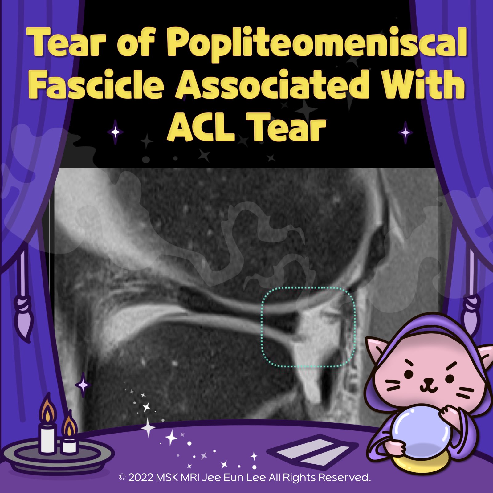
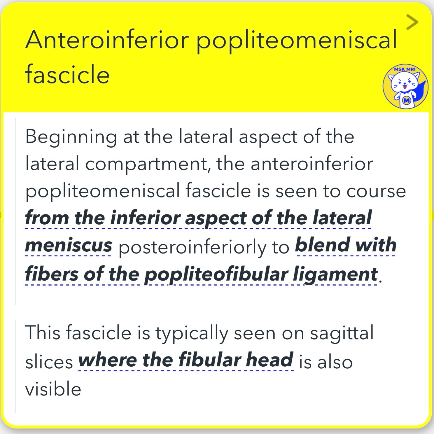
1️⃣Anteroinferior Popliteomeniscal Fascicle: Originating at the lateral compartment's lateral aspect, this fascicle runs from the inferior aspect of the lateral meniscus posteroinferiorly and merges with the popliteofibular ligament fibers.
2️⃣Posterosuperior Popliteomeniscal Fascicle: Positioned more medially, it extends from the superior margin of the lateral meniscus's peripheral posterior horn, attaching to the posterior joint capsule above the popliteus tendon.
3️⃣Posteroinferior Popliteomeniscal Fascicle: This is the least consistently visualized fascicle. When it is visible, it can be found near the root of the posterior horn, traveling from the meniscus's inferior margin posteroinferiorly to attach to a capsular extension of the popliteus tendon.
🔴🇰🇷Disruption of the popliteomeniscal fascicles
- A disruption of the popliteomeniscal fascicles can lead to increased motion of the meniscus at the hiatus, particularly causing hypermobility of the posterior horn of the lateral meniscus.
- Specifically, a disrupted posterosuperior fascicle is often associated with a tear in the posterior horn of the lateral meniscus.
"Visualizing MSK Radiology: A Practical Guide to Radiology Mastery"
© 2022 MSK MRI Jee Eun Lee All Rights Reserved.
#VisualizingMSK #ACLinjuries #Meniscocapsularseparation #PopliteomeniscalFascicles


'✅ Knee MRI Mastery > Chap 1. Meniscus' 카테고리의 다른 글
| (Fig 1-B.50) Medial Meniscocapsular Separation (0) | 2024.02.07 |
|---|---|
| (Fig 1-E.49) Isolated Tear of popliteomeniscal fascicles (0) | 2024.02.07 |
| (Fig 1-B.47) Lateral meniscocapsular separation associated with fracture (0) | 2024.02.07 |
| (Fig 1-B.46) Radial tear sparing the MFL attachment with displacement (1) | 2024.02.07 |
| (Fig 1-B.45) Radial tear sparing the MFL attachment without displacement (0) | 2024.02.07 |