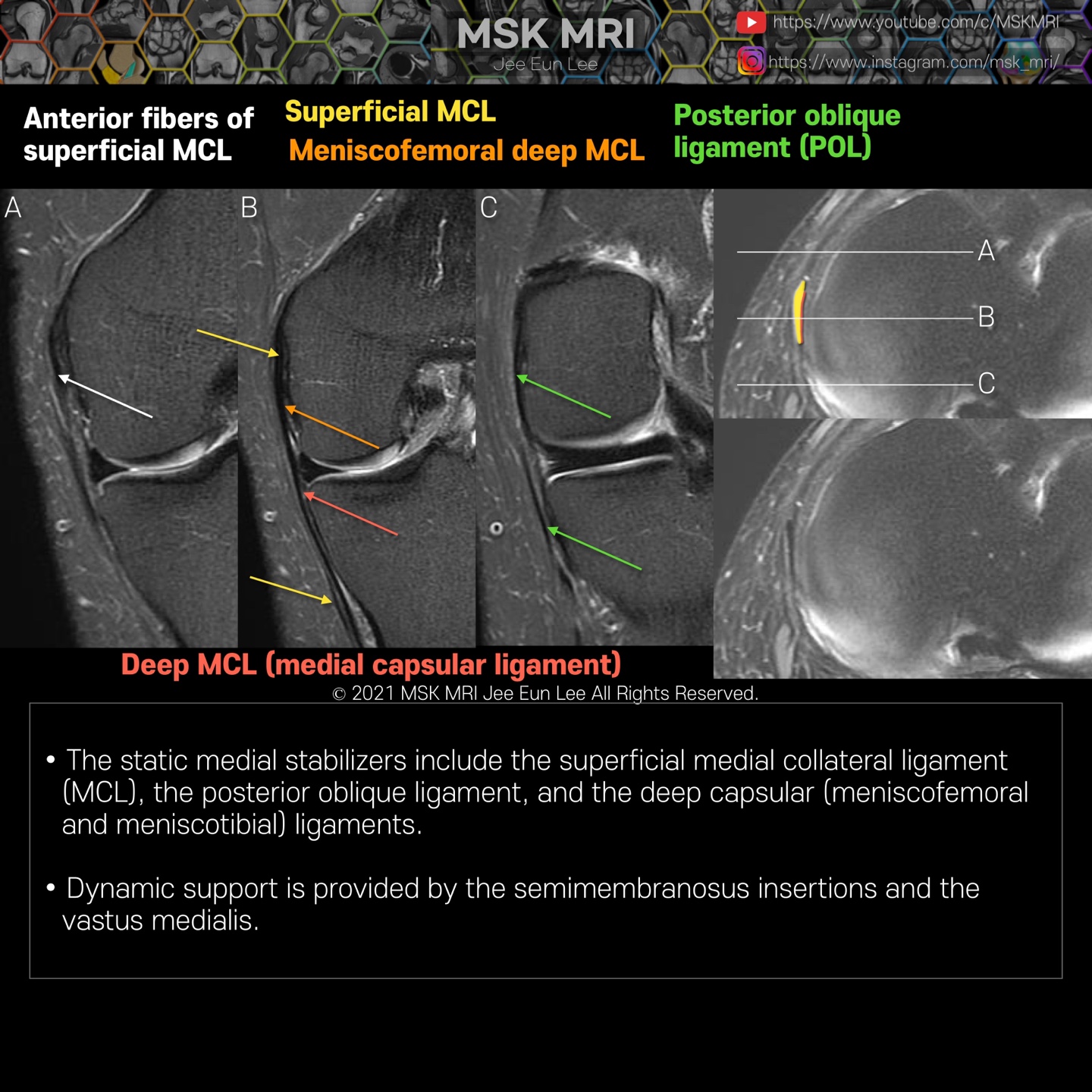
The static medial stabilizers include the superficial medial collateral ligament (MCL), the posterior oblique ligament, and the deep capsular (meniscofemoral and meniscotibial) ligaments.
Dynamic support is provided by the semimembranosus insertions and the vastus medialis.
[Axial image] The MCL has an average thickness of 4.3mm at the femoral attachment and 2.3mm at the tibial attachment.
The mean length of the dMCL attachment on the medial meniscus was 14.8 +- 3.2 mm
The anterior edges of the dMCL and sMCL are parallel and close to each other.
Posterior edge of dMCL blends with the posterior edge of sMCL.
The posteromedial corner represents structures posterior to this junction.
[Coronal, anterior]
Tears of the MCL may be overestimated if they are assessed along its anterior border. The MCL should be assessed more posteriorly.
[Coronal,middle]
The superficial MCL is 8 to 11cm long and 1.0 to 1.5cm wide. The distal attachment of the MCL is 5 to 7cm distal to the joint line on the anteromedial tibia deep to the pes insertion.
Medial capsular ligament or dMCL is composed of meniscofemoral and meniscotibial attachments to the meniscus and firmly attached to the periphery of the medial meniscus at the joint line.
The deep medial collateral ligament fuses with the superficial medial collateral ligament proximally but can be separated from the superficial MCL distally.
The femoral attachment is immediately distal to the epicondylar attachment of the sMCL
The tibial attachment is to the medial aspect of the medial rim of the tibial plateau near the joint line and proximal to the attachment of the anterior arm of the semimembranosus expansion.
[Coronal, posterior]
The MCL is attributed to primary restraint of abduction and external rotatory loads. In contrast, the posterior oblique ligament represents a secondary restraint to abduction loads, as do the cruciate ligaments.
It's not a real patient's MRI, but they are virtual images very similar to the images in the journals. The images will be created for educational purposes.
All copyrights belong to MSK MRI Jee Eun Lee.
You may not distribute or commercially exploit the content. Nor may you transmit it or store it on any other website or other forms of the electronic retrieval system.
If you would like to use an image or video for anything other than personal use, please contact me. (jamaisvu1977@gmail.com)
#Virtual MRI, #MRI illustrator, #MSKMRI © 2021 MSK MRI Jee Eun Lee All Rights Reserved.


#MSKMRI, #virtualMRI, #radiologist, #Knee_MRI, #MSKMRI_Knee, #Knee_anatomy, #Knee_meniscus, #meniscus, #Virtual_MRI, #MRI_illustrator, #posteriorobliqueligament, #posteriorcapsule, #medialcollateralligament, #MCL,
