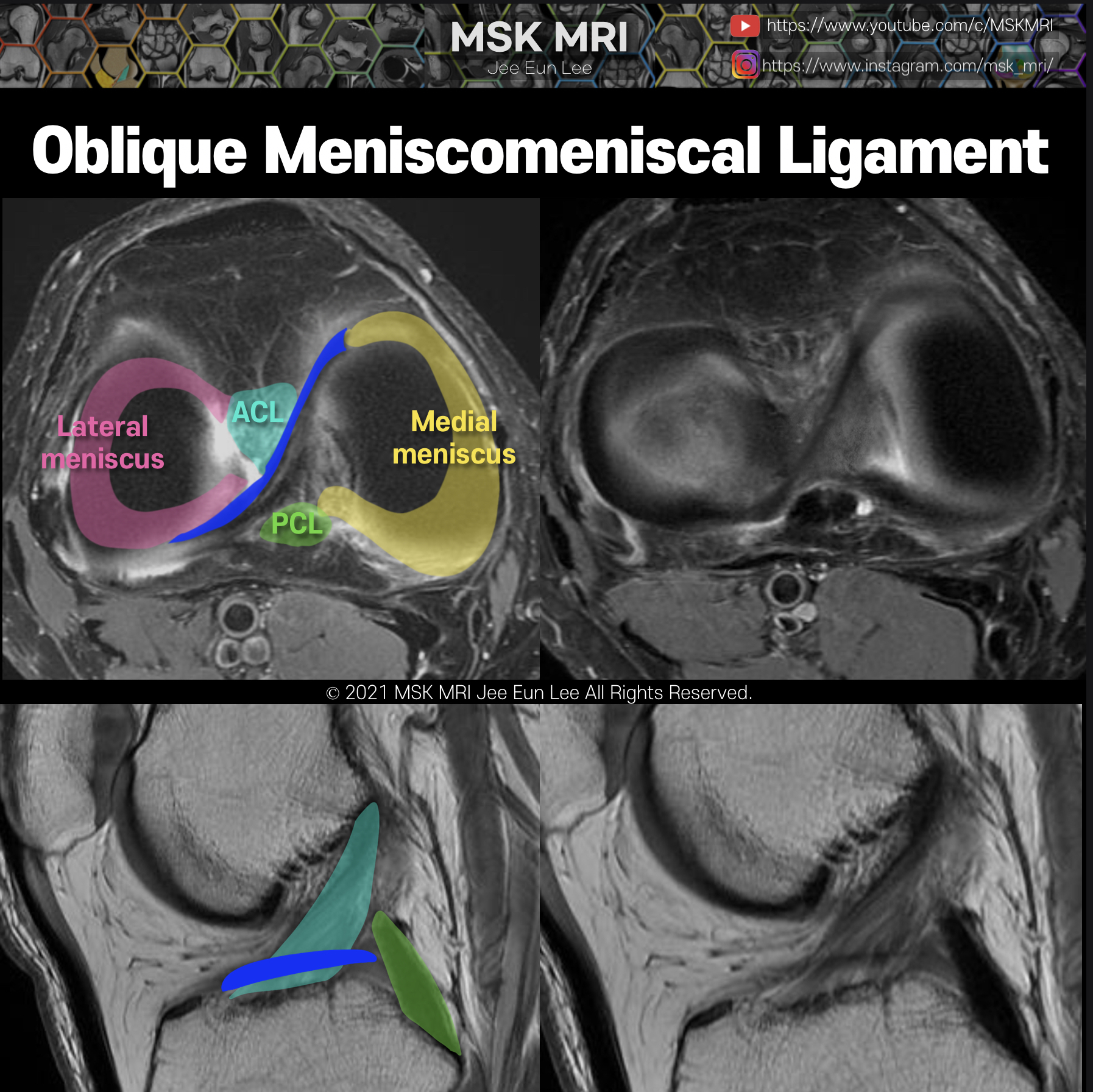
The medial OMML originates from the anterior horn of the medial meniscus and extends through the intercondylar notch to insert onto the posterior horn of the lateral meniscus.
This ligament passes between the ACL and PCL.
This oblique meniscomeniscal ligament is kind of similar SI to meniscal tissue.
The sagittal image near the midline demonstrates a linear low signal intensity structure atop the tibial plateau.
Based on this image alone, the oblique meniscomeniscal ligament may mimic a displaced meniscal fragment.
We should be careful not to call this displaced meniscal tear.


© 2021 MSK MRI Jee Eun Lee All Rights Reserved.
You may not distribute or commercially exploit the content. Nor may you transmit it or store it on any other website or other forms of the electronic retrieval system.
If you would like to use an image or video for anything other than personal use, please contact me.
(jamaisvu1977@gmail.com)
#MSKMRI, #virtualMRI, #radiologist, #Knee_MRI, #MSKMRI_Knee, #Knee_anatomy, #Knee_meniscus, #meniscus, #Virtual_MRI, #MRI_illustrator,
#bucket_handle_tear, #Oblique_Meniscomeniscal_Ligament, #meniscaltear, #meniscustear
'Knee MRI > Meniscus' 카테고리의 다른 글
| [Tear_03] Longitudinal Meniscal Tear -01 (0) | 2021.10.09 |
|---|---|
| [Anatomy_30] Oblique Meniscomeniscal Ligament vs bucket handle tear -03 (0) | 2021.10.02 |
| [Anatomy_28] Oblique Meniscomeniscal Ligament -01 (2) | 2021.10.02 |
| [Anatomy_27] Anterior transverse meniscal ligament -02 (0) | 2021.09.27 |
| [Anatomy_26] Anterior transverse meniscal ligament -01 (0) | 2021.09.27 |