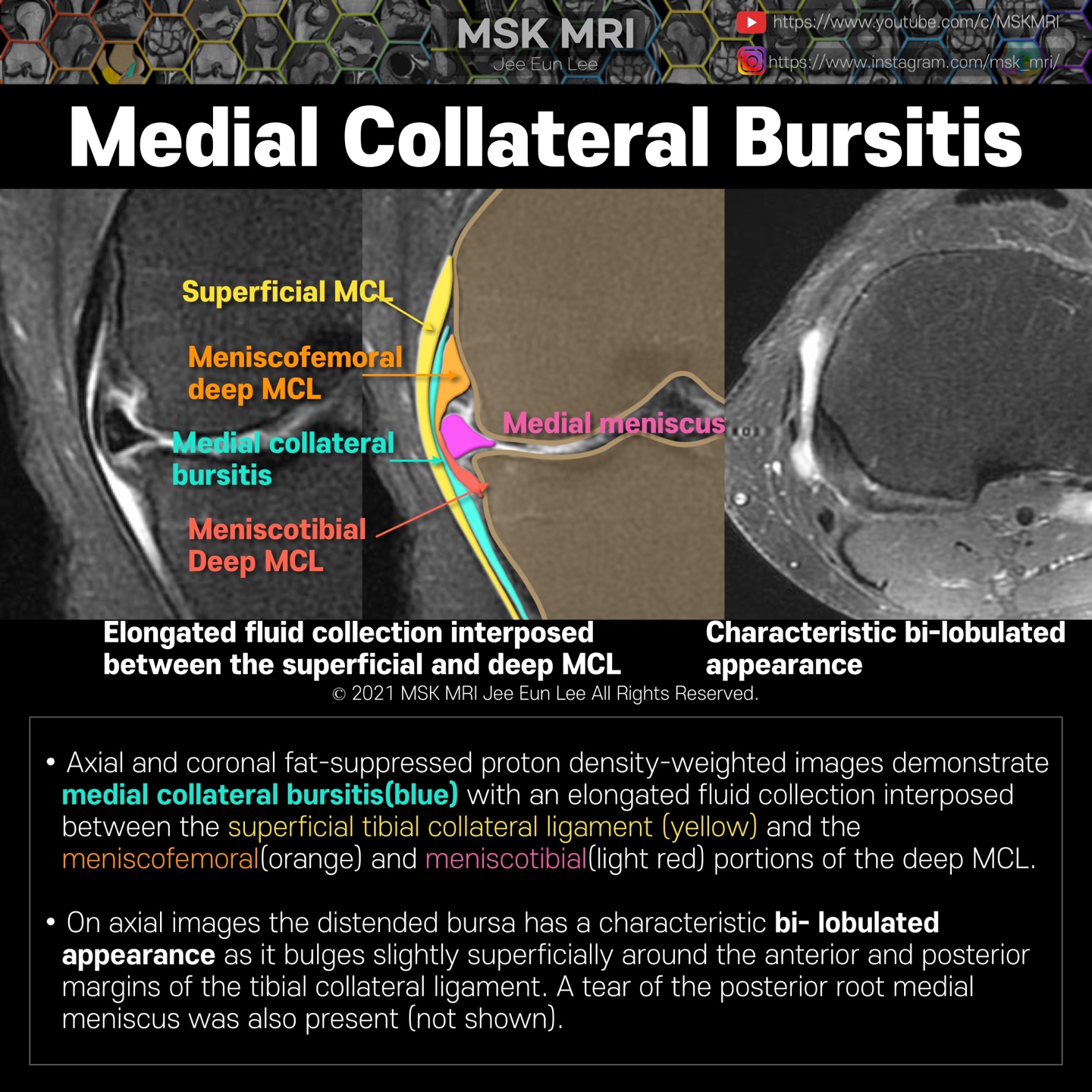
https://youtu.be/ZCOejlDr3xw Axial and coronal fat-suppressed proton density-weighted images demonstrate medial collateral bursitis(blue) with an elongated fluid collection interposed between the superficial tibial collateral ligament (yellow) and the meniscofemoral(orange) and meniscotibial(pink) portions of the deep MCL. On axial images, the distended bursa has a characteristic bi- lobulated a..