https://youtube.com/shorts/19TwSM3CQRU
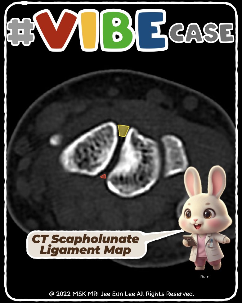
Scapholunate Ligament:
1. Three Distinct Components
The SLL has dorsal, volar, and proximal (membranous) segments, each with unique structure and function.
2. Dorsal Component – The Key Stabilizer
- Thickest and strongest portion
- Short, bandlike, and low signal on MRI
- Controls translation and resists scaphoid flexion
- Most commonly completely torn in symptomatic instability
3. Volar Component – Rotation Controller
- Much thinner (≤1 mm) with oblique collagen fibers
- Controls segmental rotation and contributes to proprioception
- Often shows striated, heterogeneous increased signal (normal)
- Must not be confused with the overlying radiolunotriquetral (RLT) ligament
4. Proximal/Membranous Component – Weakest Segment
- Fibrocartilaginous and meniscus-like
- Wedge- or triangular-shaped on imaging
- Inserts on hyaline cartilage rather than cortical bone
- Common site of degenerative perforation
- Minimal role in stability
5. Important MRI Pitfall
The RLT ligament lies intimately over the volar SLL, so careful distinction is required to avoid falsely diagnosing a volar SLL tear.
#SLL, #scapholunate, #wristMRI, #carpalinstability, #dorsalSLL, #volarSLL, #interosseousSLL, #RLTligament, #MSKradiology, #wristanatomy, #Vibecase
Visualizing MSK Radiology: A Practical Guide to Radiology Mastery
© 2022 MSK MRI Jee Eun Lee All Rights Reserved.
No unauthorized reproduction, redistribution, or use for AI training.


'✅ Dr. Slothic Notes' 카테고리의 다른 글
| 📌 Fibrous Dysplasia on MRI_ What’s the pattern? 👀 (0) | 2025.12.12 |
|---|---|
| 📌 Fibrous Dysplasia on Radiograph & CT — Classic Features to Recognize (0) | 2025.12.12 |
| 📌 Scapholunate Ligament: Gross Anatomy and Key MRI Distinctions (0) | 2025.12.10 |
| 📌 CT-Based Predictors of Stability in Lumbar Pars Fractures (0) | 2025.12.08 |
| 📌 Incomplete Spondylolysis: The Early Pars Fracture We Must Not Miss (0) | 2025.12.07 |