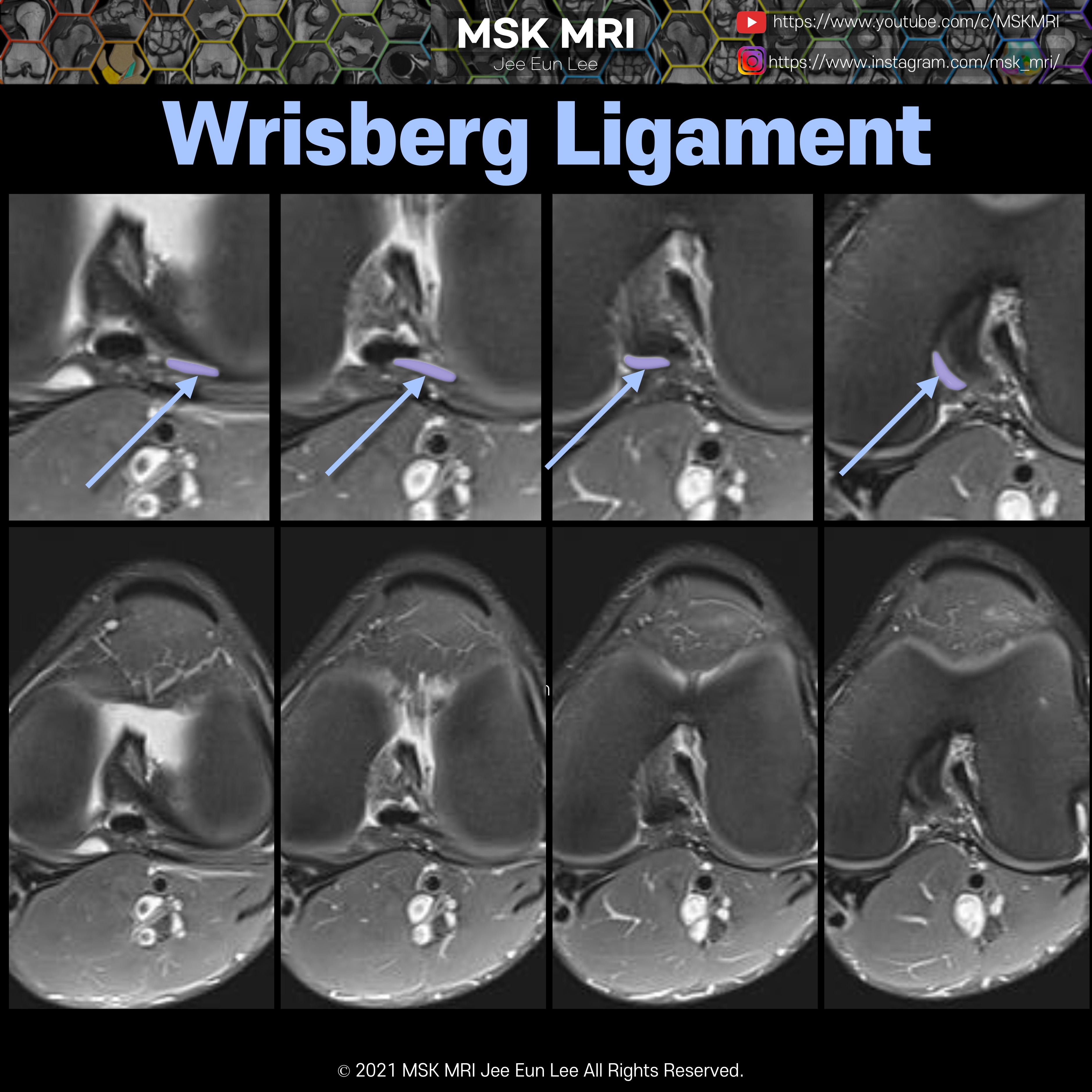
Coronal image demonstrates the posterior meniscofemoral ligament of Wrisberg (arrow) coursing obliquely downward from the medial femoral condyle to attach to the posterior horn of the lateral meniscus
The meniscofemoral ligaments originate from the posterior horn of the lateral meniscus and insert onto the lateral aspect of the posterior medial femoral condyle, posterior to the PCL.
notice, This cleft between the meniscus and meniscofemoral ligament may be mistaken for a meniscal tear.
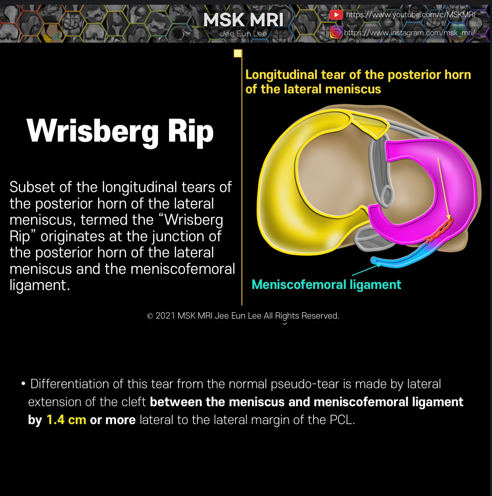
Subset of the longitudinal tears of the posterior horn of the lateral meniscus, termed the “Wrisberg Rip” originates at the junction of the posterior horn of the lateral meniscus and the meniscofemoral ligament
Differentiation of this tear from the normal pseudo-tear is made by lateral extension of the cleft between the meniscus and meniscofemoral ligament by 1.4 cm or more lateral to the lateral margin of the PCL.
Apparent far lateral meniscal extension of a meniscofemoral ligament should be considered as a possible PHLM tear, especially in the setting of an ACL tear


© 2021 MSK MRI Jee Eun Lee All Rights Reserved.
#MSKMRI, #virtualMRI, #radiologist, #Knee_MRI, #MSKMRI_Knee, #Knee_anatomy, #Knee_meniscus, #meniscus, #Virtual_MRI, #MRI_illustrator, #lateralmeniscus, #LM, #popliteomeniscalfascicle, #lateralmeniscustear, #Humphry, #Wrisberg, #Wrisbergrip
'Knee MRI > Meniscus' 카테고리의 다른 글
| [Anatomy_25] Coronary (meniscotibial) ligament of lateral meniscus (0) | 2021.09.26 |
|---|---|
| [Anatomy_24] Meniscofemoral ligament, Humphry ligament (0) | 2021.09.26 |
| [Shorts #05] Meniscofemoral ligament, Wrisberg ligament #shorts (0) | 2021.09.26 |
| [Anatomy_22] Meniscofemoral ligament, Humphry, Wrisberg (0) | 2021.09.26 |
| [Shorts #03] Meniscofemoral ligament, Humphry, Wrisberg #shorts (0) | 2021.09.25 |