Peripheral attachments of the lateral meniscus are less extensive and generally more lax than on the medial side , allowing greater mobility of the lateral meniscus, and likely contributing to the relatively increased frequency of medial meniscal tears.
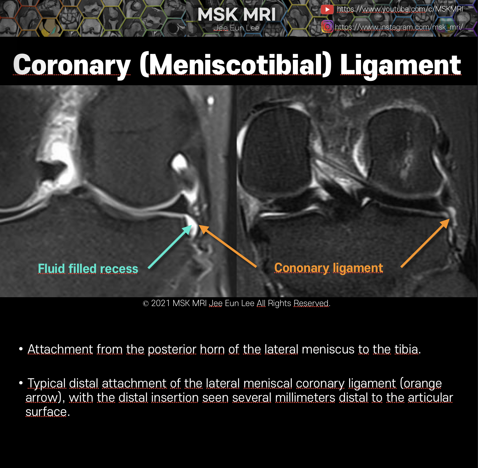
The coronary ligaments (meniscotibial ligaments) provide an attachment from the posterior horn of the lateral meniscus to the tibia.
This fat-suppressed proton density-weighted coronal image demonstrates the typical distal attachment of the lateral meniscal coronary ligament (arrow), with the distal insertion seen several millimeters distal to the articular surface. not the level of just articular surface
Because of the distal attachment of the coronary ligaments, this structure may result in visualization of a fluid filled recess as on this posterior coronal image.
© 2021 MSK MRI Jee Eun Lee All Rights Reserved.
You may not distribute or commercially exploit the content. Nor may you transmit it or store it on any other website or other forms of the electronic retrieval system.
If you would like to use an image or video for anything other than personal use, please contact me.
(jamaisvu1977@gmail.com)


#MSKMRI, #virtualMRI, #radiologist, #Knee_MRI, #MSKMRI_Knee, #Knee_anatomy, #Knee_meniscus, #meniscus, #Virtual_MRI, #MRI_illustrator, #lateralmeniscus, #LM, #lateralmeniscustear, #coronaryligament, #meniscotibialligament, #meniscotibial,
'Knee MRI > Meniscus' 카테고리의 다른 글
| [Anatomy_27] Anterior transverse meniscal ligament -02 (1) | 2021.09.27 |
|---|---|
| [Anatomy_26] Anterior transverse meniscal ligament -01 (0) | 2021.09.27 |
| [Anatomy_24] Meniscofemoral ligament, Humphry ligament (0) | 2021.09.26 |
| [Anatomy_23] Meniscofemoral ligament, Wrisberg ligament, PseudoTear, Wrisberg Ri (0) | 2021.09.26 |
| [Shorts #05] Meniscofemoral ligament, Wrisberg ligament #shorts (0) | 2021.09.26 |