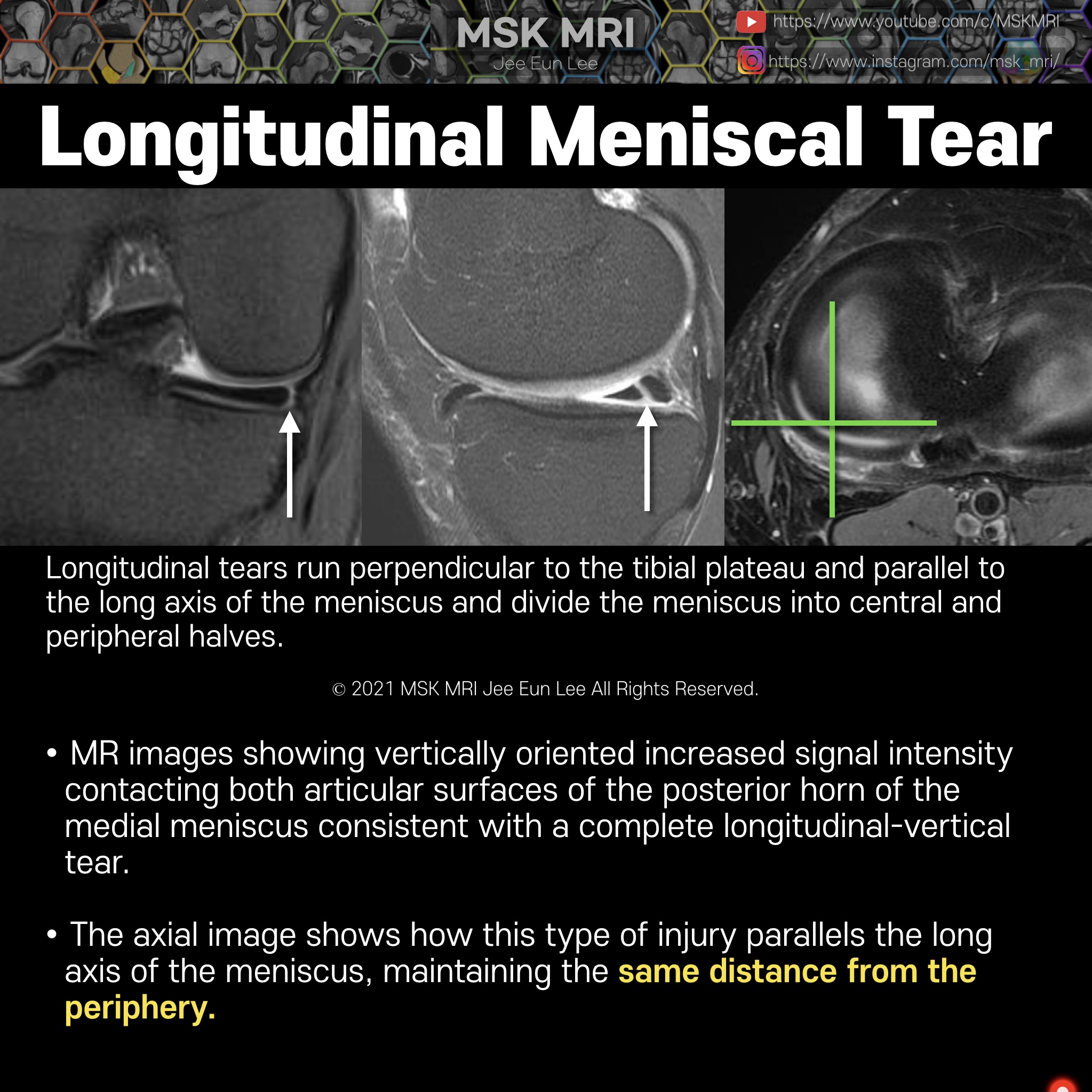
Longitudinal tears almost always involve the posterior horn in both the medial and lateral menisci.
They are diagnosed on MRI by the presence of a vertical line of increased signal intensity contacting the superior, inferior, or both surfaces of the meniscus.
MR images showing vertically oriented increased signal intensity contacting both articular surfaces of the body and posterior horn of the medial meniscus consistent with a complete longitudinal-vertical tear.
The axial image shows how this type of injury parallels the long axis of the meniscus, maintaining the same distance from the periphery.
This type of tear should not extend to the free edge.


© 2021 MSK MRI Jee Eun Lee All Rights Reserved.
You may not distribute or commercially exploit the content. Nor may you transmit it or store it on any other website or other forms of the electronic retrieval system.
If you would like to use an image or video for anything other than personal use, please contact me.
(jamaisvu1977@gmail.com)
#MSKMRI, #virtualMRI, #radiologist, #Knee_MRI, #MSKMRI_Knee, #Knee_meniscus, #meniscus, #Virtual_MRI, #MRI_illustrator, #Meniscustear,
#Longitudinaltear,
'Knee MRI > Meniscus' 카테고리의 다른 글
| [Tear_06] Far Peripheral Tear Longitudinal Meniscal Tear -04 (0) | 2021.10.09 |
|---|---|
| [Tear_05] Inferior Surface Longitudinal Meniscal Tear -03 (0) | 2021.10.09 |
| [Tear_03] Longitudinal Meniscal Tear -01 (0) | 2021.10.09 |
| [Anatomy_30] Oblique Meniscomeniscal Ligament vs bucket handle tear -03 (0) | 2021.10.02 |
| [Anatomy_29] Oblique Meniscomeniscal Ligament -02 (0) | 2021.10.02 |