Meniscal ramp lesions can be defined as longitudinal vertical and/or oblique peripheral tears in the posterior horn of medial meniscus, in a mediolateral direction of less than 2.0 cm, that may lead to meniscocapsular or meniscotibial disruption with a concomitant ACL injury.
There are few classifications regarding meniscal ramp lesions.
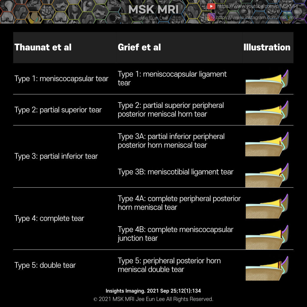
Thaunat et al. approached a more comprehensive classification system which incorporates tear pattern, direction, tear thickness (partial versus full), and associated meniscocapsular junction, red-red zone, or meniscotibial ligament disruption and instability.
Greif et al. extended Thaunat classification.
Let's summarize the ramp lesion using Grief classification.
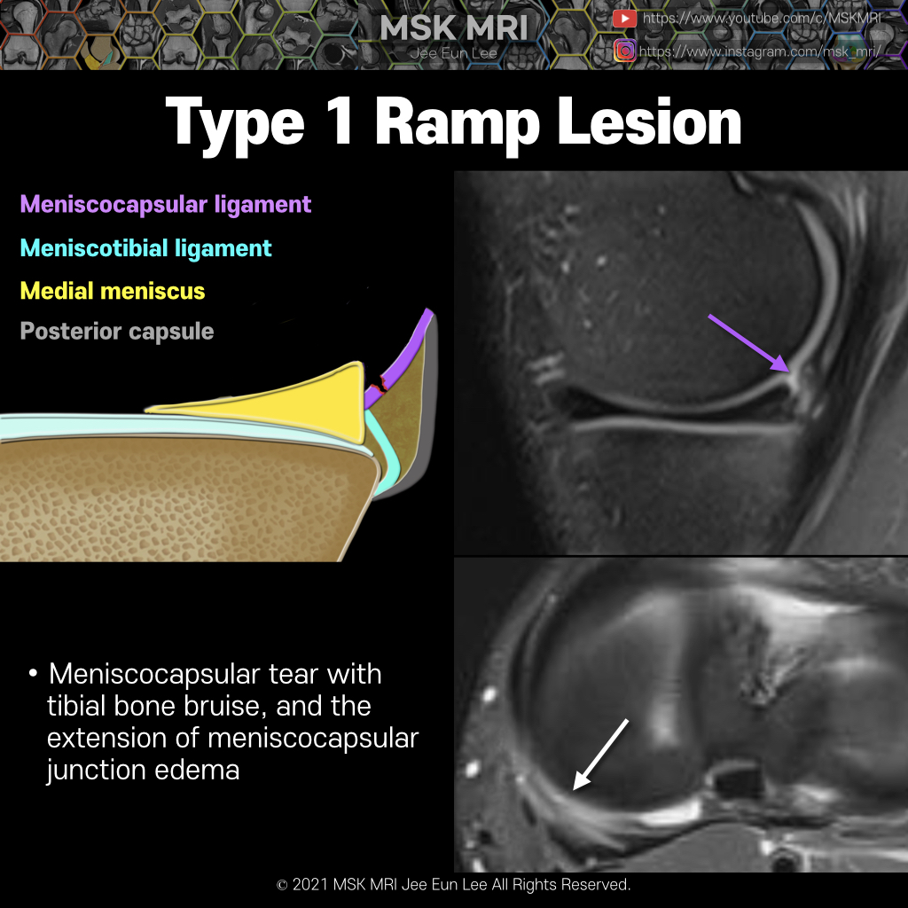
Type 1 Ramp lesion is an isolated posterior superior meniscocapsular tear.
Meniscocapsular tears are located peripherally and involve the synovium, leading to posterior meniscocapsular separation from the posterior horn of the medial meniscus.
Ramp lesion type 2 is a peripheral partial thickness tear involving the superior margin of the posterior horn, where the torn fragment contains the intact meniscocapsular attachment to the posterior horn.
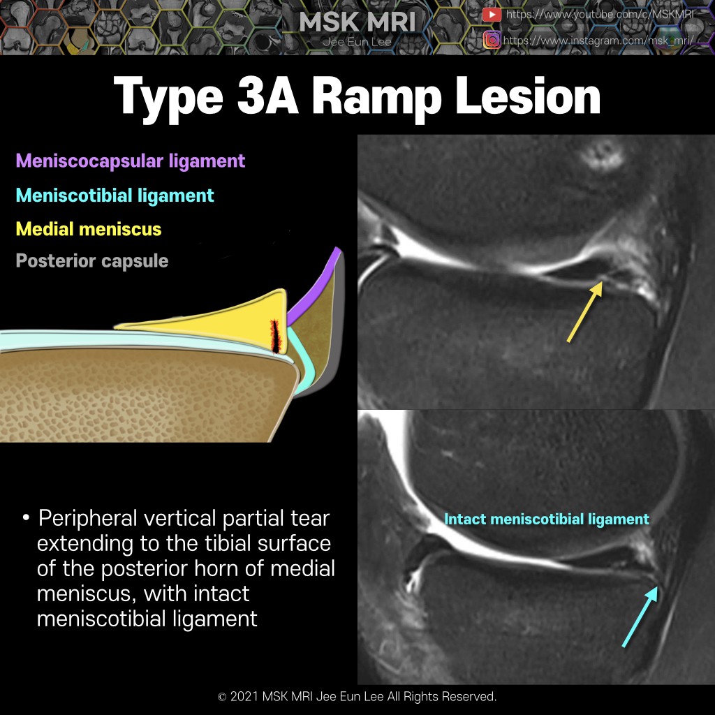
Type 3A represents a vertical peripheral tear of the inferior margin of the posterior horn containing the attachment of the meniscotibial ligament.
Though not torn, the meniscotibial ligament is no longer directly connected to the medial meniscus, and thus becomes unstable.
The type 3B is a tear of the meniscotibial ligament itself from its attachment to the posterior horn.
This includes rupture in the midsubstance of the ligament or an avulsion of the ligament from the meniscal insertion.
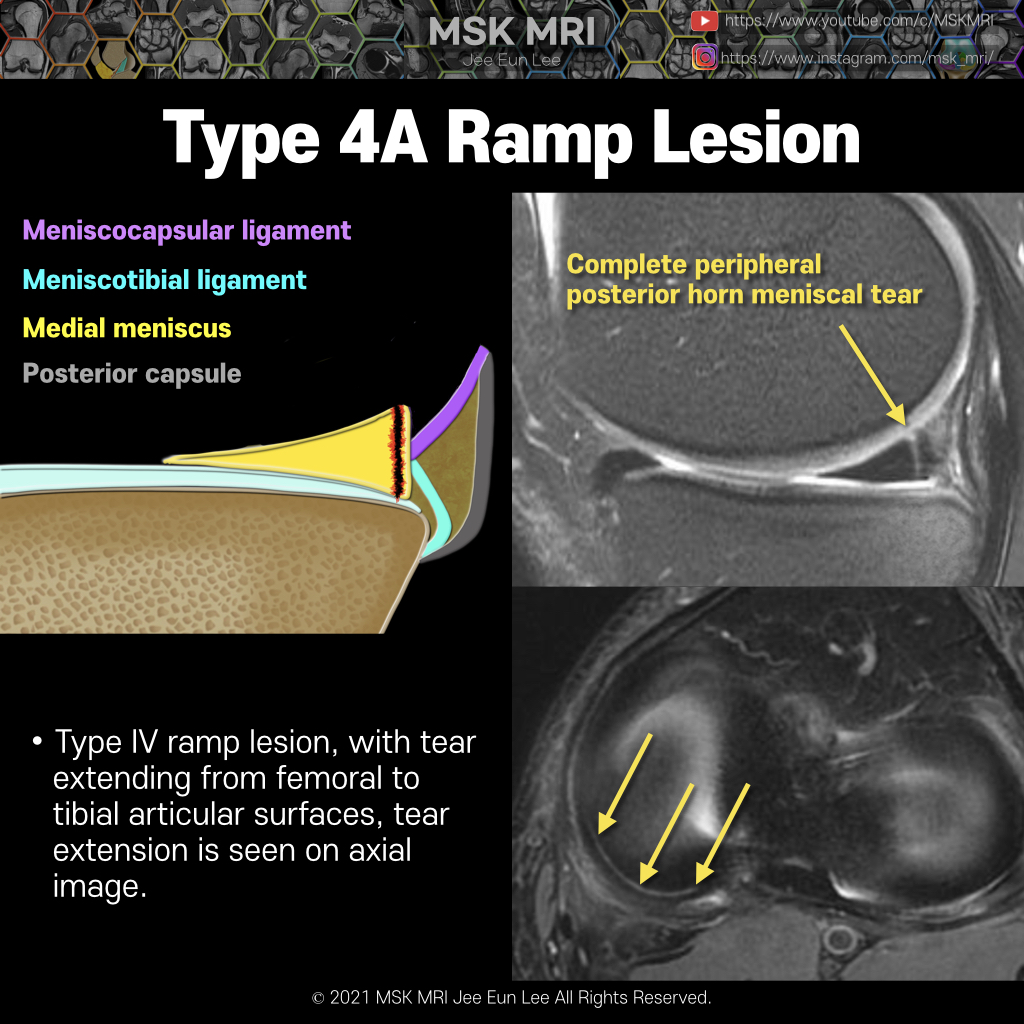
The subtype 4A is a complete longitudinal vertical tear of the red-red zone of the meniscus, with the meniscocapsular and meniscotibial ligaments intact but connected to a portion of the posterior horn junction via a free-floating fragment of the meniscus, thus leading to instability of both structures
The subtype 4B involves a complete tear of the junction itself where the meniscocapsular and meniscotibial fibers attach to the posterior horn.
Edema and irregularity of the meniscocapsular junction of the posterior horn are the most frequent findings.
A fluid signal cleft at the meniscocapsular junction is most specific for making the diagnosis.
In type5 tear pattern, there are two separate tears within the red- red zone of the meniscus. So this is called a double tear.
Type 5 ramp lesions are similar to type 4a.
Similar to the 4A subtype, the meniscocapsular and meniscotibial ligaments are intact.


© 2021 MSK MRI Jee Eun Lee All Rights Reserved.
You may not distribute or commercially exploit the content. Nor may you transmit it or store it on any other website or other forms of the electronic retrieval system.
If you would like to use an image or video for anything other than personal use, please contact me.
(jamaisvu1977@gmail.com)

#MSKMRI, #virtualMRI, #radiologist, #Knee_MRI, #MSKMRI_Knee, #Knee_anatomy, #Knee_meniscus, #meniscus, #Virtual_MRI, #MRI_illustrator, #medialmeniscus, #MM, #meniscustear, #medialmeniscustear, #Ramplesion, #longitudinaltear, #ACLtear,#meniscocapsularseparation