Stage I
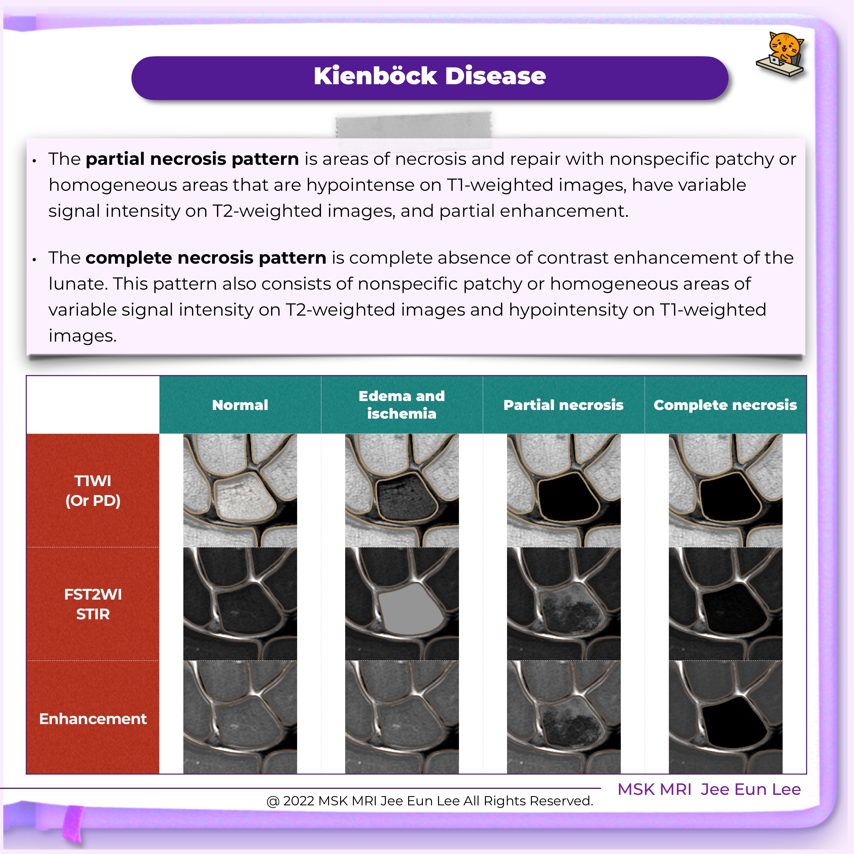
• Bone marrow edema due to ischemic damage without morphological changes
• Normal density of the lunate in radiography and attenuation at CT scan
• Low signal intensity on T1-weighted images and usually hyperintense on T2-weighted fat-suppressed images.
Stage II
• Sclerosis of the lunate bone due to chronic ischemic damage
• Increased density in radiography with preserved morphology
• CT is more accurate in determining subtle fracture lines and pseudo-cystic inclusions
• Low signal intensity on T1-weighted and variable signal intensity on T2-weighted images.
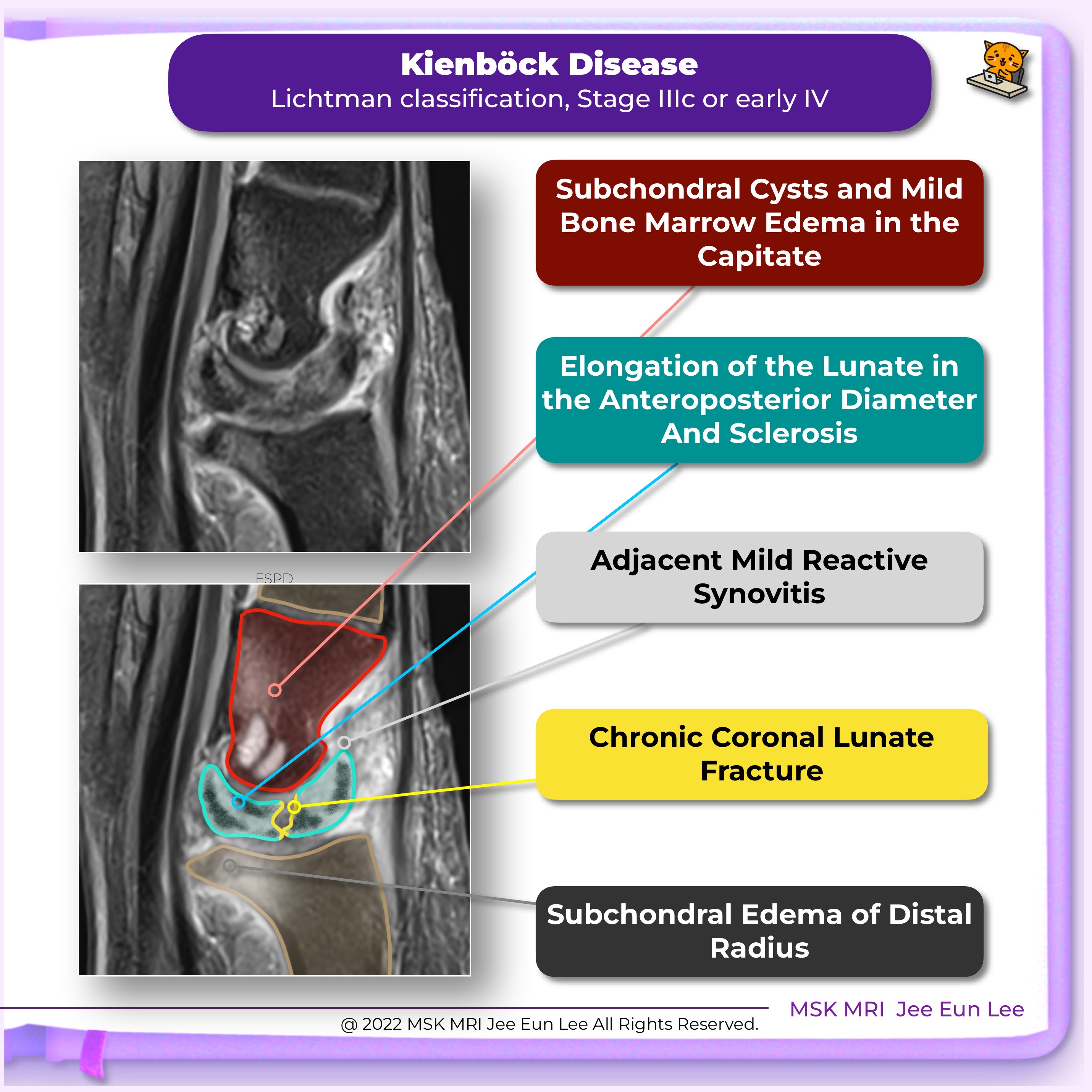
Stage III
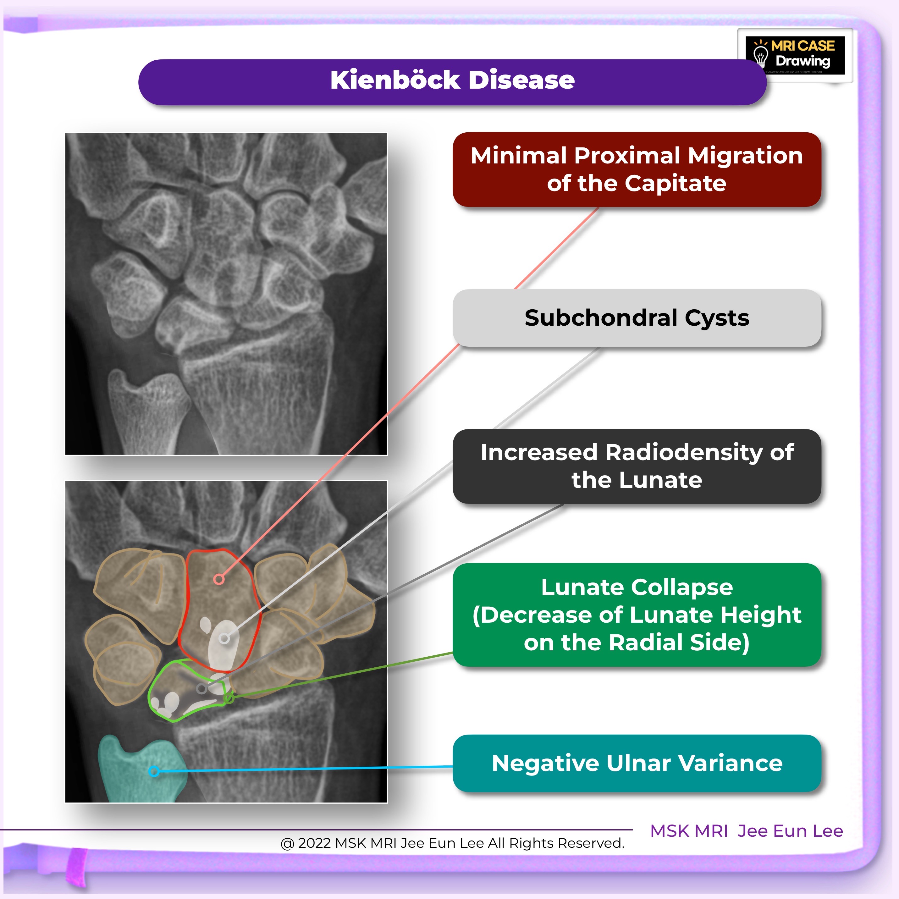
• Sclerosis and collapse of the lunate
• No signs of degenerative arthritis changes in the other wrist bones
• Increased density in radiography and collapse of the lunate
• CT reveals fractures, deformity, and widening on the sagittal plane
• Low signal intensity on T1-weighted images and variable signal intensity on T2-weighted images.
Three subgroups
• Stage IIIa - no carpal instability (radio-scaphoid angle <60°)
• Stage IIIb - carpal instability (radio-scaphoid angle >60°)
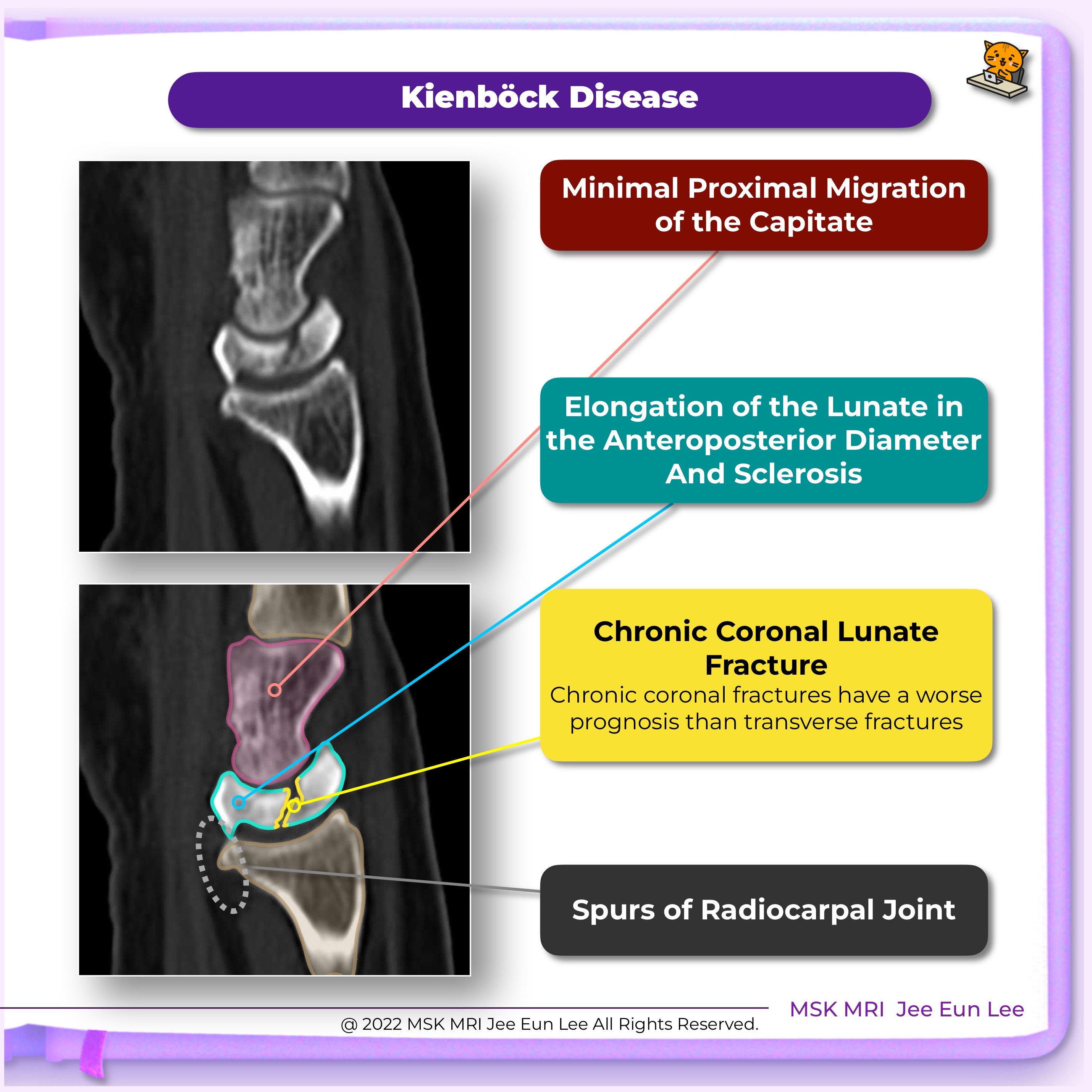
• Stage IIIc - chronic coronal fractures of the lunate
Stage IV
• Collapse of the lunate
• Carpal instability • Radiocarpal and midcarpal degenerative arthritis
• Functional impairment
• High density in radiography and high CT attenuation
• Low signal intensity on T1 and T2-weighted images due to sclerosis and necrosis.
💟 Visit my youtube channel for a detailed description
Youtube channel: MSKMRI
💟 Visit my Instagram: msk_mri
https://www.instagram.com/msk_mri/
🥰 #followme,
#WristMRI, #Kienböck's disease, #Kienböckdisease, #lunate, #lunatecollapse, #osteonecrosis,
#안산에이스병원,#Ansan_Ace_Hospital,#MSK_MRI, #mskmri_wrist, #영상의학과이지은, #공부맛집, #영상의학공부맛집, #studygram,