| Category | Description |
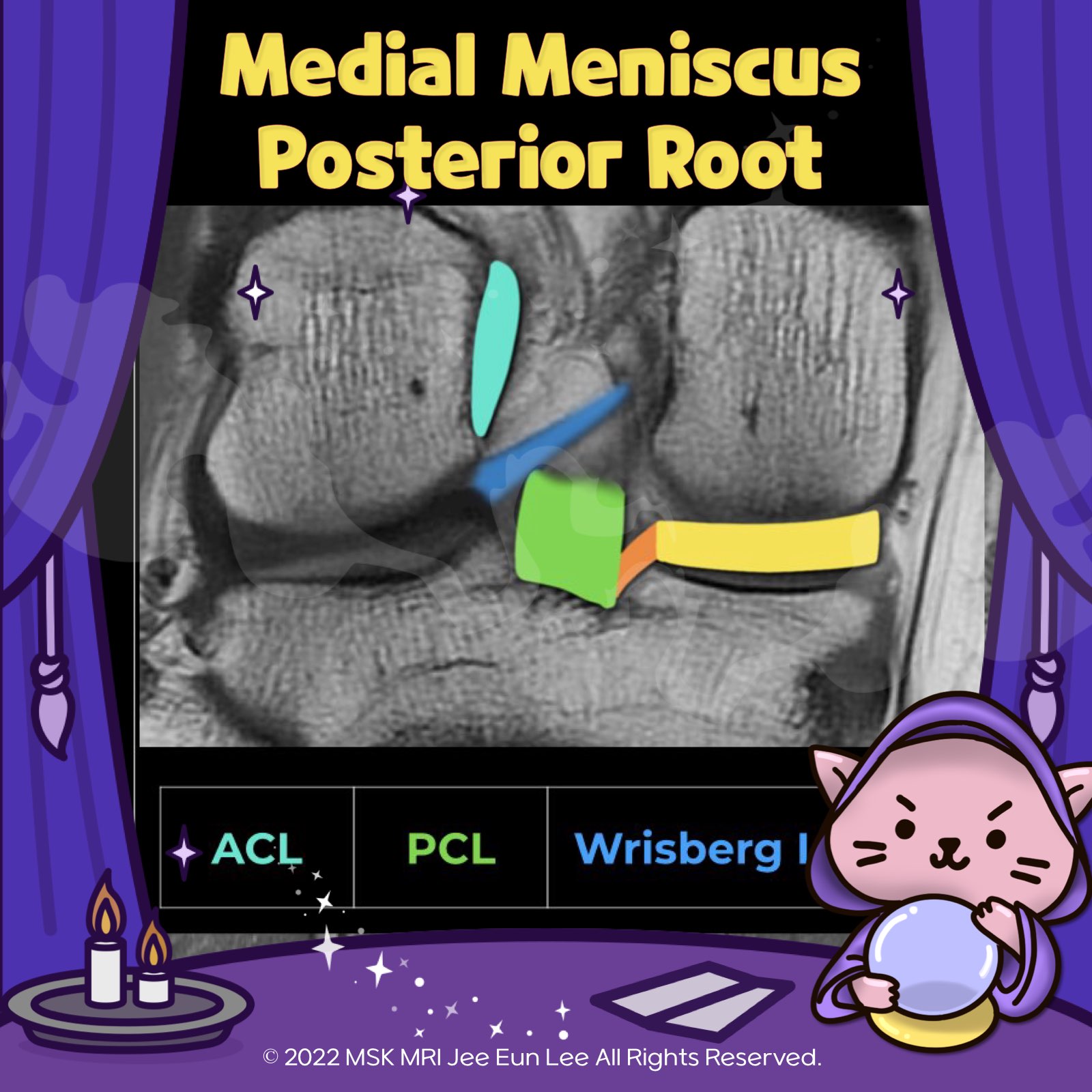
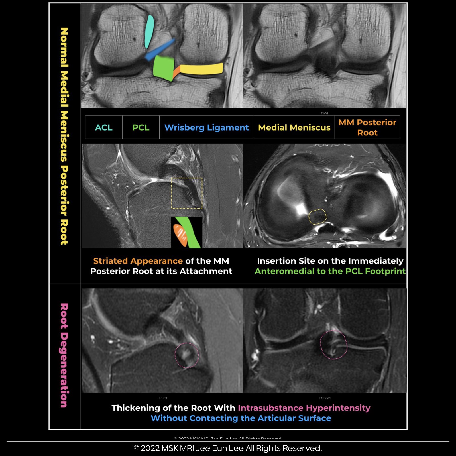
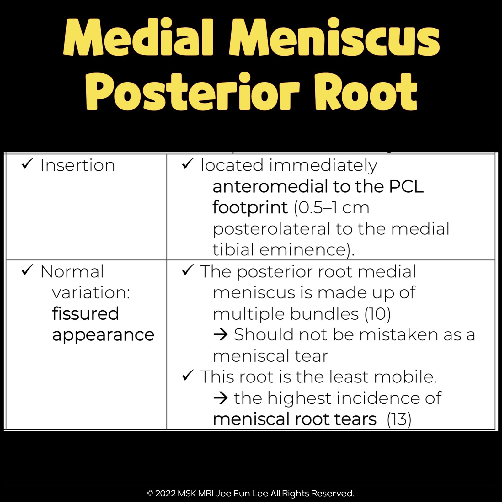
| Insertion Points | - Inserts along the downward slope of the intercondylar fossa, anterior to the posterior cruciate ligament. - Extends from the tibial attachment point to just lateral from the articular cartilage inflection point of the medial tibial plateau. - Commences just lateral to the apex of the medial tibial plateau. |
| Function | - Anterior and posterior meniscal roots help resist hoop stress >- Prevent outward displacement of the meniscus during axial loading. |
| Composition | - Composed of multiple bundles. |
| Variations | - May have a fissured appearance, a normal variation that should not be mistaken for a meniscal tear. |
| Lesion Locations | - Enthesial Location: At the root attachment of the ligament to the underlying tibia. - Midsubstance Location: Within the root ligament. - Junctional Location: At the attachment of the ligament with the posterior horn. |
© 2021 MSK MRI Jee Eun Lee All Rights Reserved.
You may not distribute or commercially exploit the content.
Nor may you transmit it or store it on any other website or other forms of the electronic retrieval system.
If you would like to use an image or video for anything other than personal use, please contact me. (jamaisvu1977@gmail.com) or (jamaisvu77@naver.com)