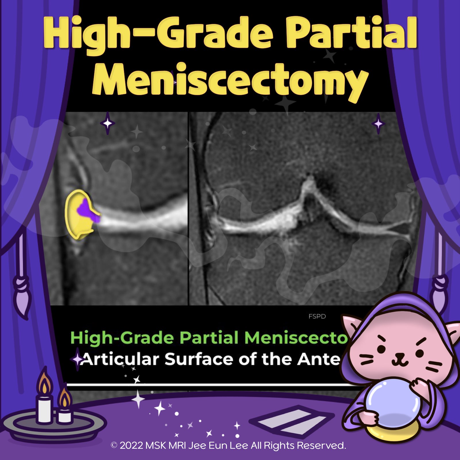
✅ High-grade partial meniscectomy (>25%)
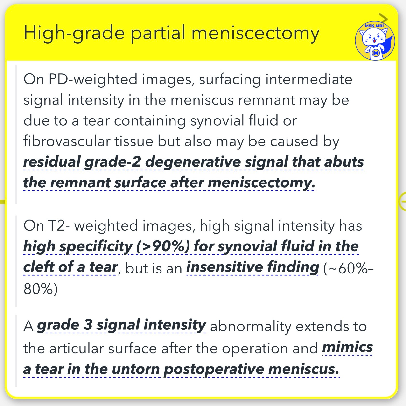
- In cases of high-grade partial meniscectomy, where more than 25% of the meniscus is removed, diagnosing tears becomes less accurate.
- For T2-weighted MRI scans, a high signal intensity specifically indicates synovial fluid within a tear's cleft with over 90% specificity, but this sign lacks sensitivity (about 60%–80%).
📌 Criteria for identifying a recurrent tear after more than 25% of the meniscus has been resected include:
- A definite surfacing T2 fluid signal (or a high T1 signal that is isointense to intra-articular gadolinium in MR arthrography) visible on two or more images or the presence of a displaced meniscal fragment.
- A definitive surfacing fluid signal on just one image suggests a possible tear.


"Visualizing MSK Radiology: A Practical Guide to Radiology Mastery"
© 2022 MSK MRI Jee Eun Lee All Rights Reserved. #VisualizingMSK #meniscaltear #meniscus #meniscectomy
You may not distribute or commercially exploit the content. Nor may you transmit it or store it on any other website or other forms of the electronic retrieval system. If you would like to use an image or video for anything other than personal use, please contact me. (jamaisvu1977@gmail.com) or (jamaisvu77@naver.com) (instagram: msk_mri)

'✅ Knee MRI Mastery > Chap 1. Meniscus' 카테고리의 다른 글
| (Fig 1-E.05) Recurrent Tear in partial meniscectomy -2 (0) | 2024.02.09 |
|---|---|
| (Fig 1-E.04) Recurrent Tear in partial meniscectomy -1 (0) | 2024.02.09 |
| (Fig 1-E.02) Expected appearance of Low-grade partial meniscectomy. (0) | 2024.02.09 |
| (Fig 1-E.01) Preferential resection of the inner margin of the inferior leaf (0) | 2024.02.09 |
| (Fig 1-D.03) Accessory iliotibial band–meniscal ligament (1) | 2024.02.09 |