==============================================
⬇️✨⬇️🎉⬇️🔥⬇️📚⬇️
Click the link to purchase on Amazon 🎉📚
==============================================
🎥 Check Out All Videos at Once! 📺
👉 Visit Visualizing MSK Blog to explore a wide range of videos! 🩻
https://visualizingmsk.blogspot.com/?view=magazine
📚 You can also find them on MSK MRI Blog and Naver Blog! 📖
https://www.instagram.com/msk_mri/
Click now to stay updated with the latest content! 🔍✨
==============================================
✅ Segond Fracture
1️⃣ Segond Fracture and Associated Injuries
- Associated Findings: Segond fractures are linked to ACL tears, MCL tears, medial meniscus tears, and posterolateral corner injuries.
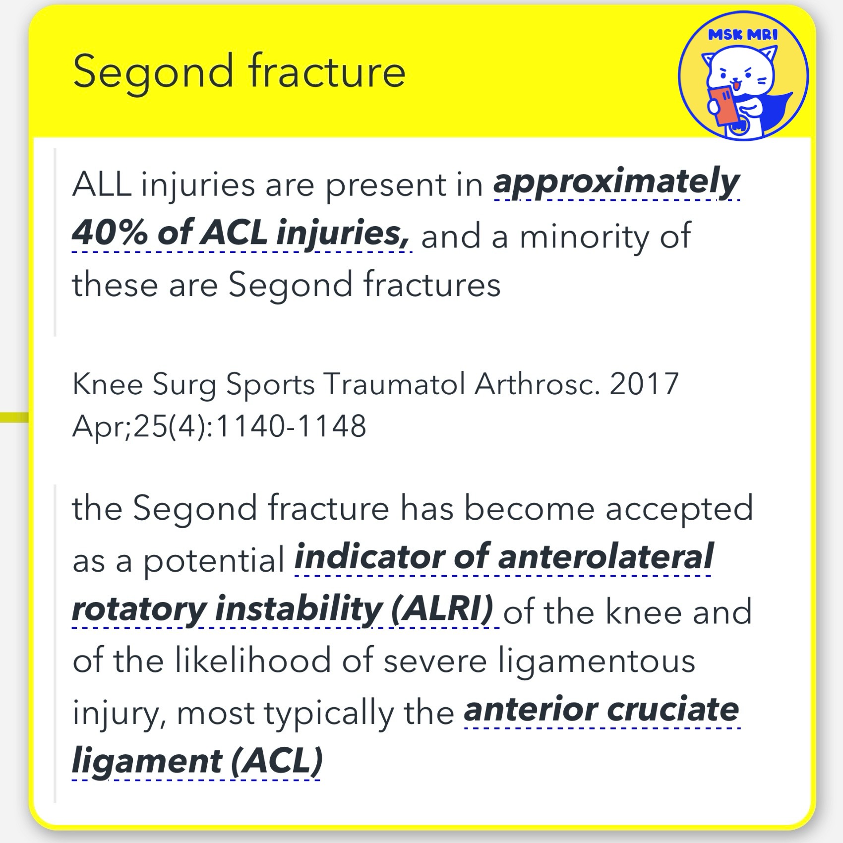
- Indicator of ALRI: Recognized as a marker for anterolateral rotatory instability (ALRI) and a predictor of severe ligamentous injury, particularly to the ACL.
2️⃣ ALL Injuries and Segond Fractures
- Prevalence in ACL Injuries: ALL injuries occur in about 40% of ACL injuries, with Segond fractures being a subset.
- Fracture Characteristics: The fracture fragment is thin, ovoid, vertically oriented, and situated anterolaterally along the lateral tibial plateau behind Gerdy’s tubercle.
3️⃣ Involvement of ITB and ALL
- Attachment to Fracture: In 94% of cases, the posterior fibers of the ITB and the lateral capsule (ALL) attach to the Segond fracture.
- Tibial Insertion of ALL: The tibial insertion of the ALL is consistently located between the fibular head and Gerdy’s tubercle and positioned between 4.0 and 7.0 mm below the tibial plateau.
Knee Surg Sports Traumatol Arthrosc. 2017 Apr;25(4):1140-1148
Am J Sports Med. 2017 Aug;45(10):2247-2252
https://visualizingmsk.blogspot.com/?view=magazine
© 2022 MSK MRI Jee Eun Lee All Rights Reserved.
#VisualizingMSK #ACLinjuries #KneeMRI #ACLtear #avulsionfracture #Segondfracture #ALRI
You may not distribute or commercially exploit the content.
Nor may you transmit it or store it on any other website or other forms of the electronic retrieval system.


'✅ Knee MRI Mastery > Chap 2.ACL and PCL' 카테고리의 다른 글
| (Fig 2-C.01) ACL Repair (0) | 2024.03.01 |
|---|---|
| (Fig 2-B.31) Ramp lesion versus no ramp lesion in ACL injury (0) | 2024.02.29 |
| (Fig 2-B.28) Avulsion fractures of the fibular head (arcuate sign) (0) | 2024.02.26 |
| (Fig 2-B.27) Scar tissue Attachment to the PCL (0) | 2024.02.25 |
| (Fig 2-B.26) Scar tissue Attachment to the Anatomical origin of the ACL (0) | 2024.02.25 |