Click the link to purchase on Amazon 🎉📚
==============================================
🎥 Check Out All Videos at Once! 📺
👉 Visit Visualizing MSK Blog to explore a wide range of videos! 🩻
https://visualizingmsk.blogspot.com/?view=magazine
📚 You can also find them on MSK MRI Blog and Naver Blog! 📖
https://www.instagram.com/msk_mri/
Click now to stay updated with the latest content! 🔍✨
==============================================
Arthrofibrosis After ACL Reconstruction
1️⃣ Focal Form: Cyclops Lesion
✅ Patients with arthrofibrosis following ACL reconstruction may present with stiffness or restricted extension, a symptom profile similar to that observed in cases of graft impingement.
✅ MRI findings
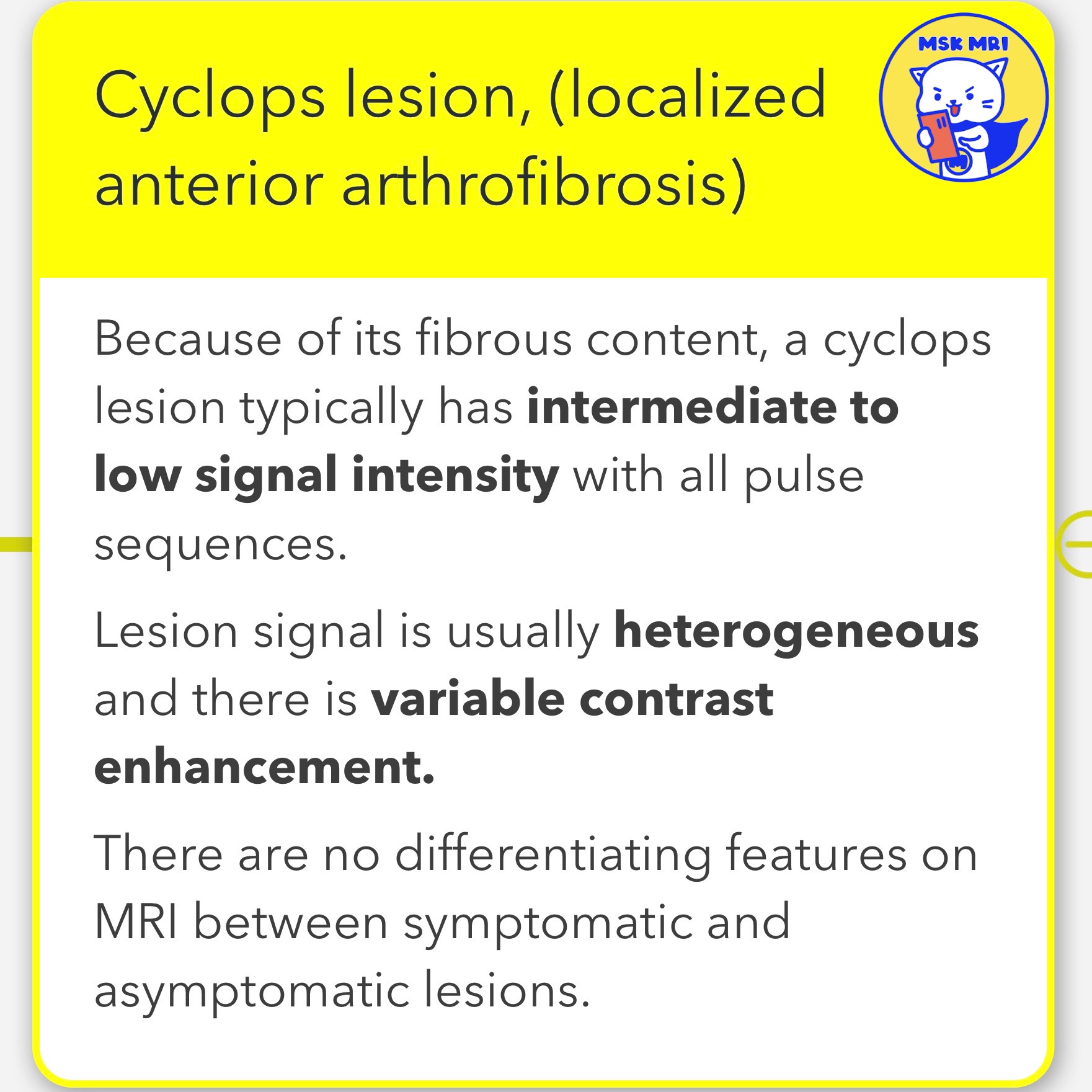
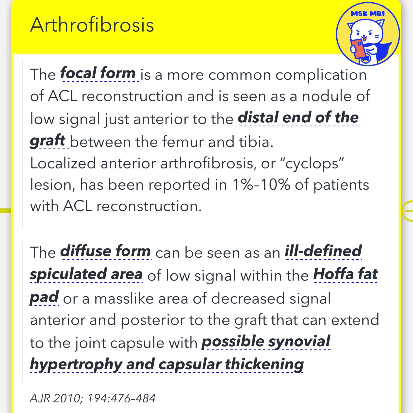
- It is characterized by a nodule of low signal intensity located just anterior to the distal end of the graft, between the femur and tibia.
- Because of its fibrous content, a cyclops lesion typically has intermediate to low signal intensity with all pulse sequences.
- Lesion signal is usually heterogeneous and there is variable contrast enhancement.
- There are no differentiating features on MRI between symptomatic and asymptomatic lesions.
2️⃣ Diffuse Form
✅MRI findings
- The diffuse form is identifiable by an ill-defined, spiculated area of low signal within the Hoffa fat pad or a mass-like area of decreased signal anterior and posterior to the graft.
- This condition can extend to the joint capsule, potentially resulting in synovial hypertrophy and capsular thickening.
Magn Reson Imaging Clin N Am. 2022 May;30(2):261-275
AJR 2010; 194:476–484
"Visualizing MSK Radiology: A Practical Guide to Radiology Mastery" © 2022 MSK MRI Jee Eun Lee All Rights Reserved. #VisualizingMSK #ACLinjuries #KneeMRI #cyclops #cyclopslesion #aclgraft You may not distribute or commercially exploit the content. You should not transmit or store it on any other website or electronic retrieval system.