Click the link to purchase on Amazon 🎉📚
==============================================
🎥 Check Out All Videos at Once! 📺
👉 Visit Visualizing MSK Blog to explore a wide range of videos! 🩻
https://visualizingmsk.blogspot.com/?view=magazine
📚 You can also find them on MSK MRI Blog and Naver Blog! 📖
https://www.instagram.com/msk_mri/
Click now to stay updated with the latest content! 🔍✨
==============================================
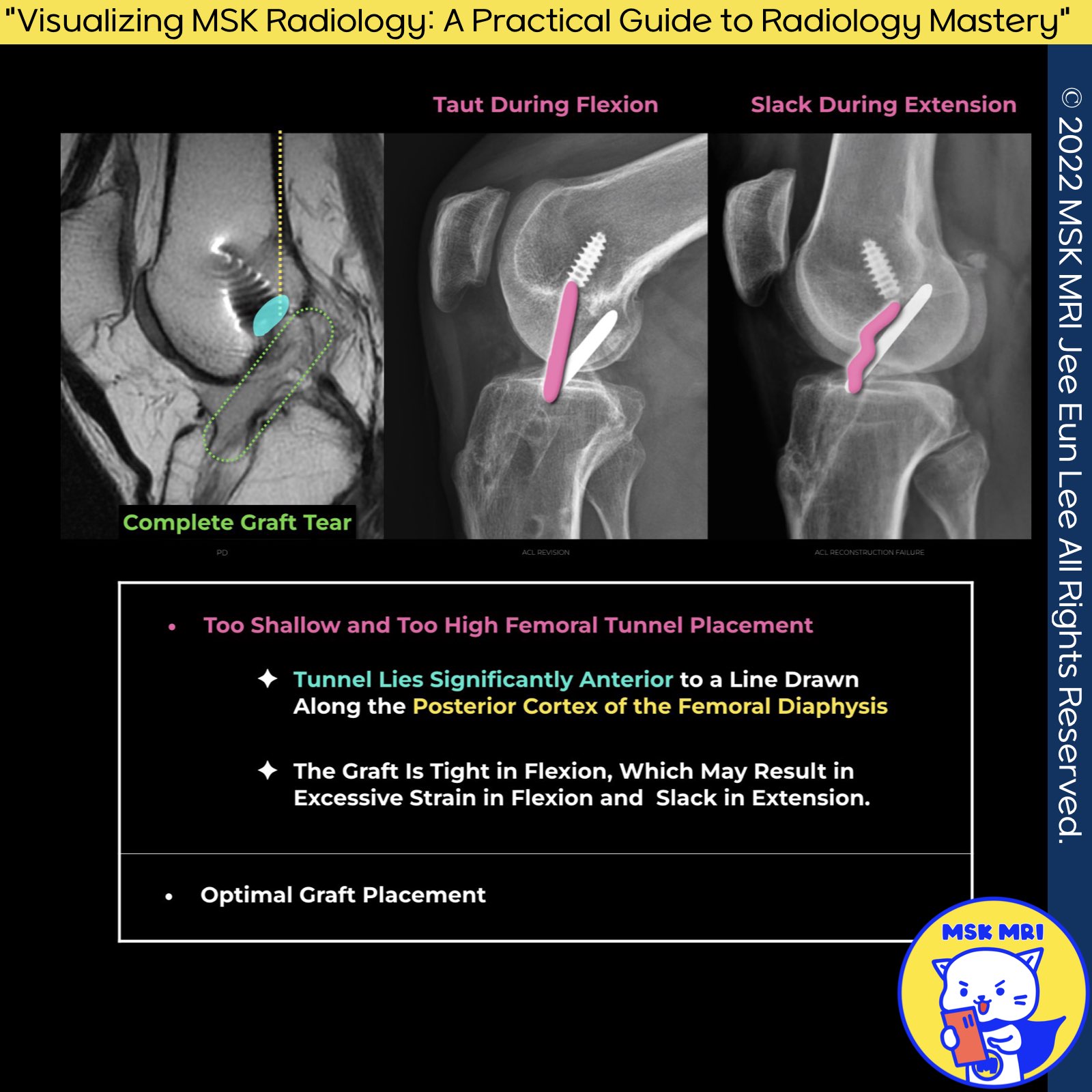
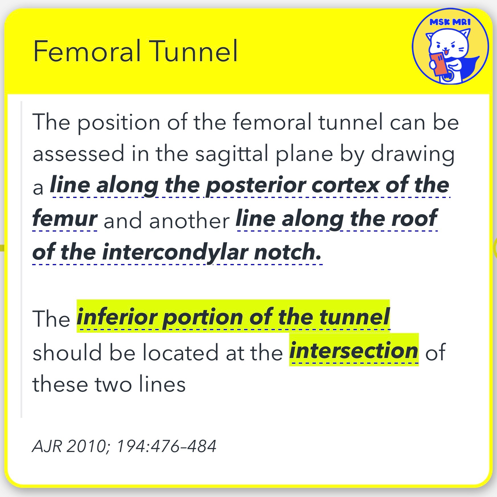
1️⃣ Assessing Femoral Tunnel Placement
✅ The position of the femoral tunnel can be assessed in the sagittal plane by drawing a line along the posterior cortex of the femur and another line along the roof of the intercondylar notch.
✅ Ideal Tunnel Location: The inferior portion of the tunnel should be located at the intersection of these two lines.
AJR 2010; 194:476–484
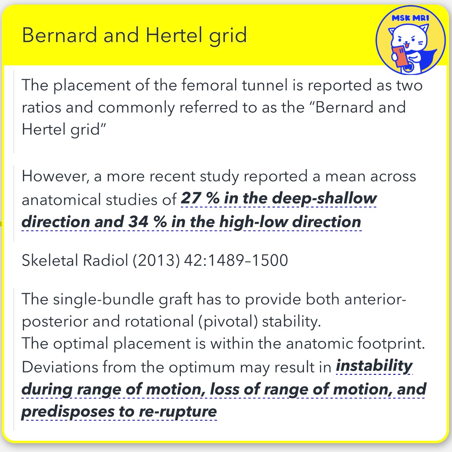
2️⃣ Bernard and Hertel Grid: Understanding Femoral Tunnel Placement
✅The femoral tunnel placement is reported as two ratios and is commonly referred to as the “Bernard and Hertel grid”.
- A more recent study reported a mean across anatomical studies of 27% in the deep-shallow direction and 34% in the high-low direction.
Skeletal Radiol (2013) 42:1489–1500
✅Importance of Single-Bundle Graft Placement
- The single-bundle graft has to provide both anterior-posterior and rotational (pivotal) stability.
- The optimal placement is within the anatomic footprint.
- Deviations from the optimum may result in instability during range of motion, loss of range of motion, and predispose to re-rupture.
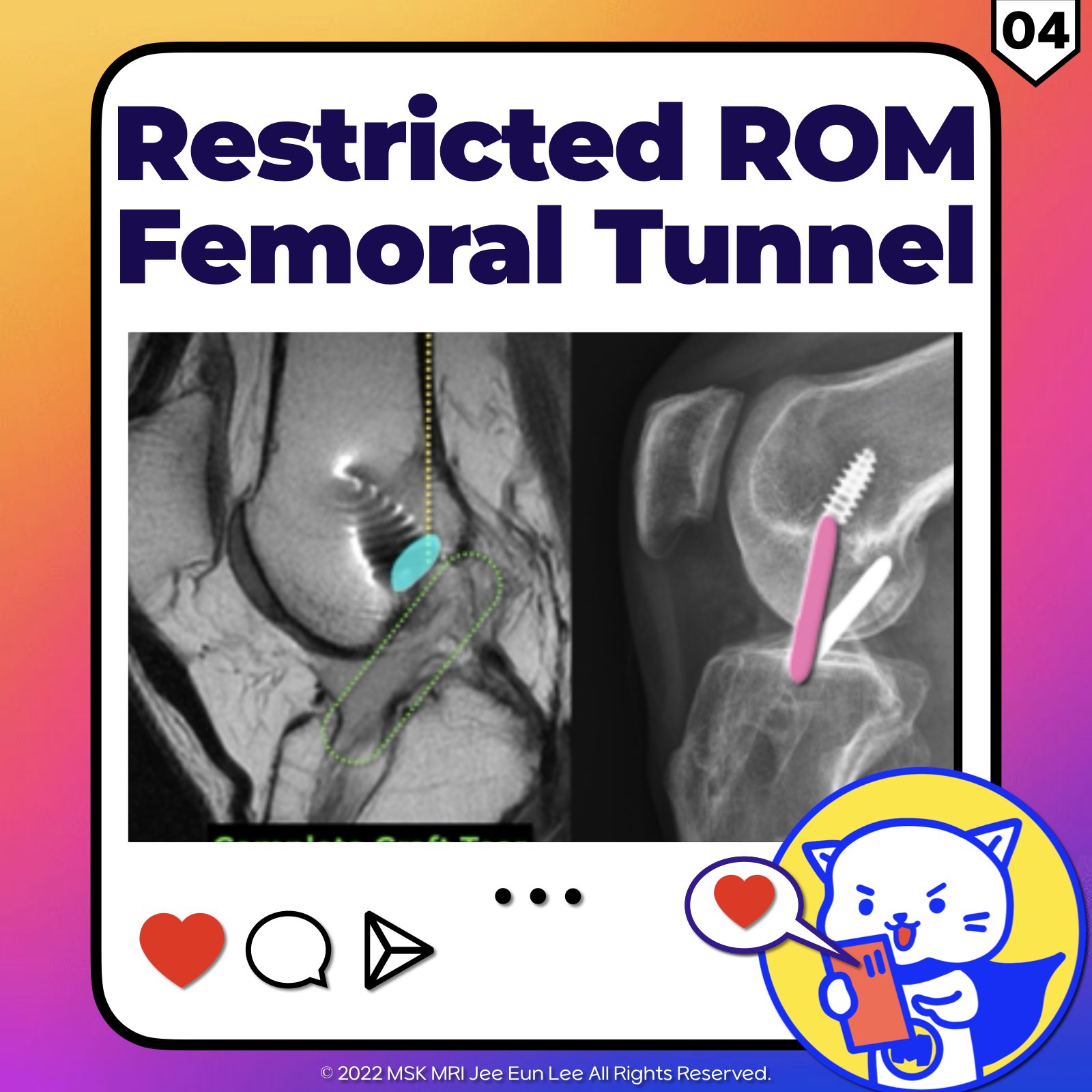
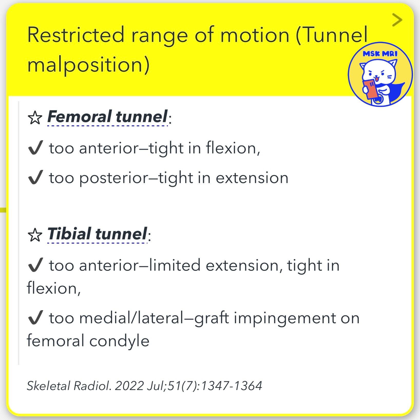
3️⃣ Restricted range of motion (Tunnel malposition)
☆ Femoral tunnel:
- ✔ too anterior—tight in flexion,
- ✔ too posterior—tight in extension
☆ Tibial tunnel:
- ✔ too anterior—limited extension, tight in flexion,
- ✔ too medial/lateral—graft impingement on femoral condyle
Skeletal Radiol. 2022 Jul;51(7):1347-1364
"Visualizing MSK Radiology: A Practical Guide to Radiology Mastery" © 2022 MSK MRI Jee Eun Lee All Rights Reserved. #VisualizingMSK #ACLinjuries #KneeMRI #ACLtear #femoraltunnel You may not distribute or commercially exploit the content. Nor may you transmit it or store it on any other website or other forms of the electronic retrieval system.
'✅ Knee MRI Mastery > Chap 2.ACL and PCL' 카테고리의 다른 글
| (Fig 2-D.06) Cyclops Lesion Arthrofibrosis After ACL Reconstruction (0) | 2024.03.09 |
|---|---|
| (Fig 2-D.05) Restricted Range of Motion Caused by Tibial Tunnel Placement, Roof impingement (0) | 2024.03.09 |
| (Fig 2-D.03) Sidewall impingement with ACL Graft tear (0) | 2024.03.09 |
| (Fig 2-D.02) Sidewall impingement ACL reconstruction (0) | 2024.03.09 |
| (Fig 2-D.01) Roof impingement (0) | 2024.03.09 |