Click the link to purchase on Amazon 🎉📚
==============================================
🎥 Check Out All Videos at Once! 📺
👉 Visit Visualizing MSK Blog to explore a wide range of videos! 🩻
https://visualizingmsk.blogspot.com/?view=magazine
📚 You can also find them on MSK MRI Blog and Naver Blog! 📖
https://www.instagram.com/msk_mri/
Click now to stay updated with the latest content! 🔍✨
==============================================
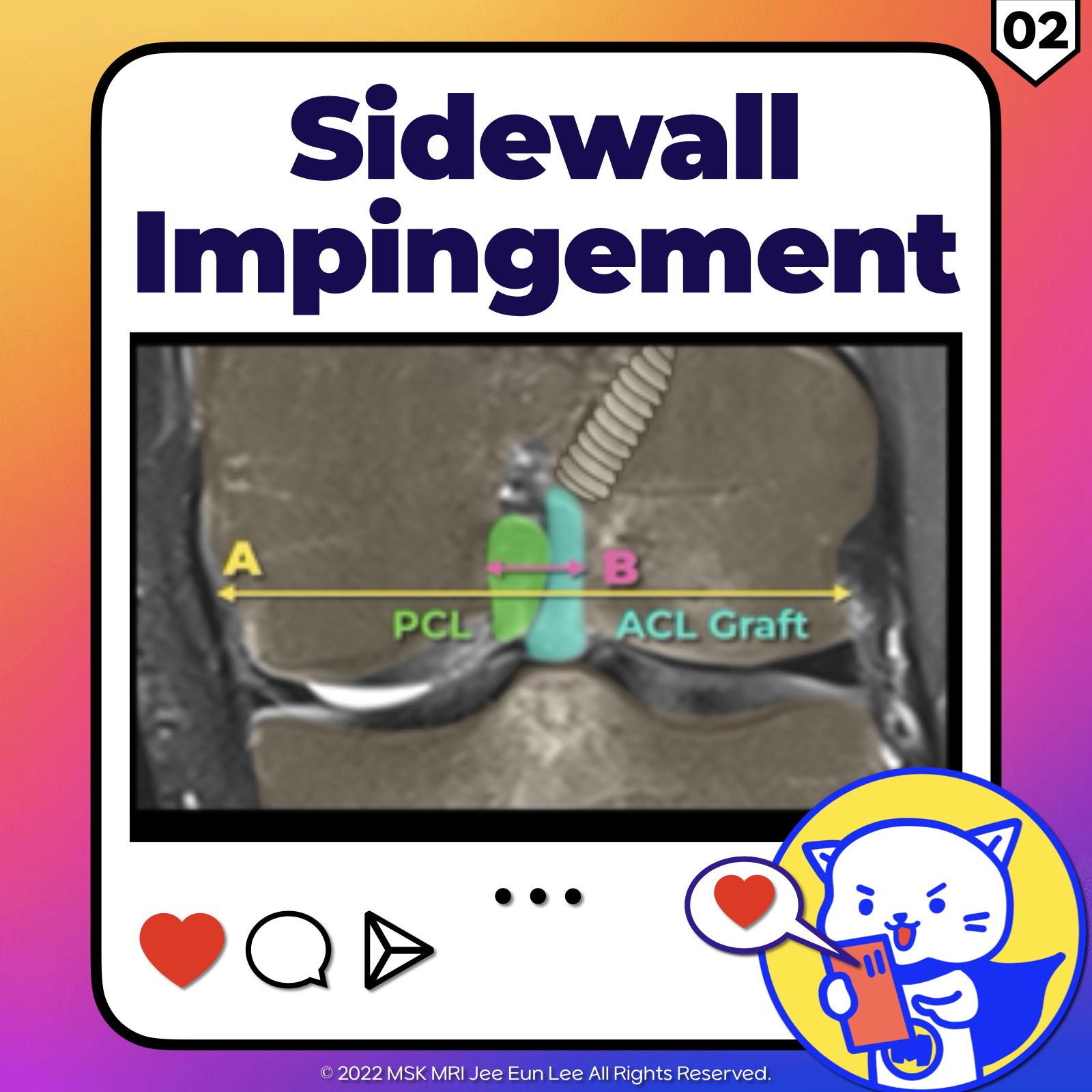
1️⃣ Side Wall Impingement
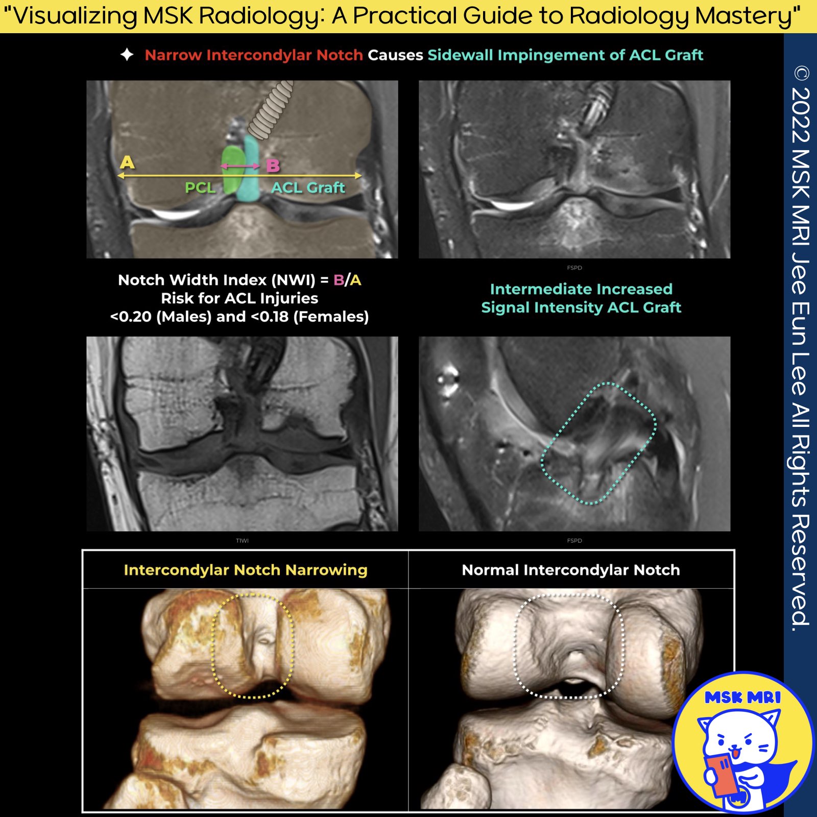
- Sidewall impingement with the intercondylar fossa is related to the shape of the fossa, graft size, and the presence of osteophytes.
- Coronal MR images show the graft indentation as it curves over the medial aspect of the lateral femoral condyle.
- This condition may be associated with the low positioning of the femoral tunnel opening in the intercondylar notch.
Stoller's Orthopaedics and Sports Medicine: The Knee
2️⃣ Notch Shape and ACL Injury Risk
- Notch shape is considered a significant risk factor for noncontact ACL injury.
- A narrow notch has been observed in ACL-deficient patients, suggesting a correlation with a smaller and weaker ACL compared to healthy patients who typically have a wider notch.
- In the context of ACL reconstruction, Fujii et al. identified a smaller notch cross-sectional area in patients who developed the "cyclops lesion" due to notch impingement.
World J Orthop. 2016 Oct 18;7(10):638-649
Knee Surg Sports Traumatol Arthrosc 2015; 23: 1092-1099
"Visualizing MSK Radiology: A Practical Guide to Radiology Mastery"
© 2022 MSK MRI Jee Eun Lee All Rights Reserved. #VisualizingMSK #Lateralmeniscus #Discoidmeniscus #Meniscaltears #Poplitealmeniscacal #Meniscalinstability

"Visualizing MSK Radiology: A Practical Guide to Radiology Mastery" © 2022 MSK MRI Jee Eun Lee All Rights Reserved. #VisualizingMSK #ACLinjuries #KneeMRI #ACLtear #aclgraft #graftimpingement You may not distribute or commercially exploit the content. Nor may you transmit it or store it on any other website or other forms of the electronic retrieval system.
'✅ Knee MRI Mastery > Chap 2.ACL and PCL' 카테고리의 다른 글
| (Fig 2-D.04) Restricted Range of Motion Caused by Femoral Tunnel (0) | 2024.03.09 |
|---|---|
| (Fig 2-D.03) Sidewall impingement with ACL Graft tear (0) | 2024.03.09 |
| (Fig 2-D.01) Roof impingement (0) | 2024.03.09 |
| (Fig 2-C.13) MRI Characteristics of Different Graft Type (0) | 2024.03.03 |
| (Fig 2-C.12) ACL Graft Ligamentization (0) | 2024.03.03 |