==============================================
⬇️✨⬇️🎉⬇️🔥⬇️📚⬇️
Click the link to purchase on Amazon 🎉📚
==============================================
🎥 Check Out All Videos at Once! 📺
👉 Visit Visualizing MSK Blog to explore a wide range of videos! 🩻
https://visualizingmsk.blogspot.com/?view=magazine
📚 You can also find them on MSK MRI Blog and Naver Blog! 📖
https://www.instagram.com/msk_mri/
Click now to stay updated with the latest content! 🔍✨
==============================================
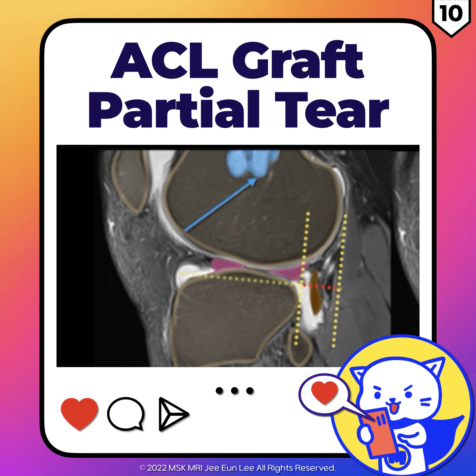
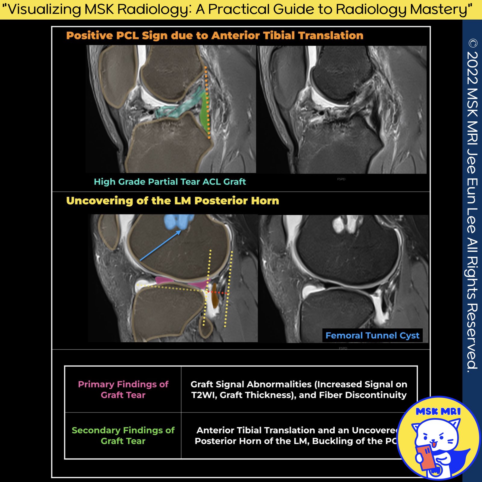
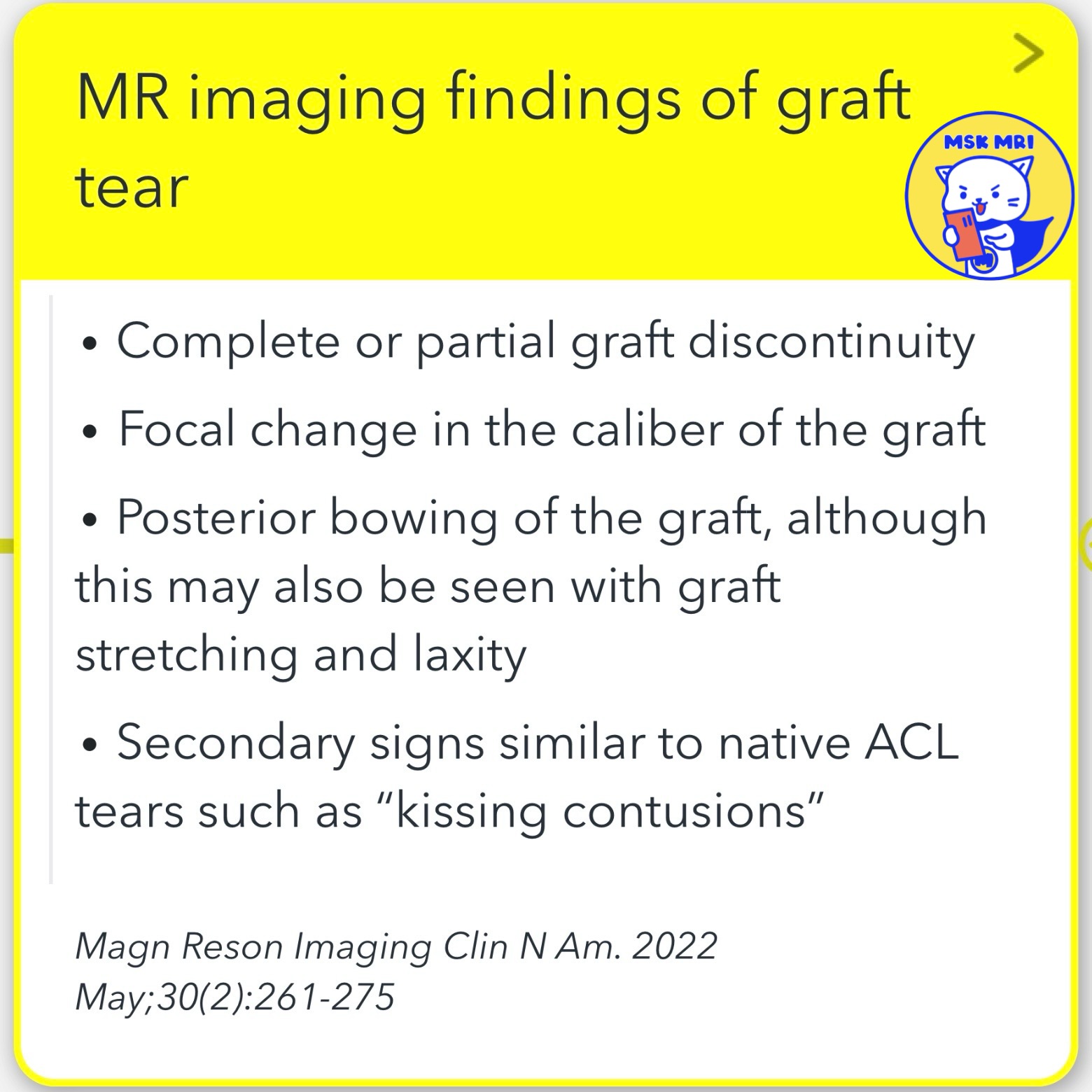
✅ Differentiating Revascularization from Graft Tear
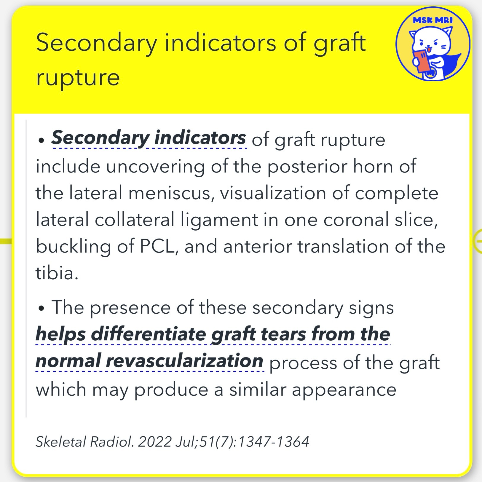
- Secondary indicators of graft rupture include uncovering the lateral meniscus's posterior horn, visualization of the complete lateral collateral ligament in one coronal slice, buckling of PCL, and anterior translation of the tibia.
- The observation of secondary signs is crucial in distinguishing between graft tears and the normal revascularization process of the graft, which might present a similar appearance on imaging.
- Clinical Signs of Instability: These, rather than a decreased range of motion, and a history of re-injury, are critical factors in differentiating a rupture from impingement or the subacute stage of ligamentization in challenging cases.
Skeletal Radiology (2022) 51:1347–1364
"Visualizing MSK Radiology: A Practical Guide to Radiology Mastery"
© 2022 MSK MRI Jee Eun Lee All Rights Reserved.
#VisualizingMSK #ACLinjuries #KneeMRI #ACLtear #aclgrafttear #ligamentization
You should not distribute or commercially exploit the content.
You should not transmit or store it on any other website or electronic retrieval system.

'✅ Knee MRI Mastery > Chap 2.ACL and PCL' 카테고리의 다른 글
| (Fig 2-D.12) Chronic ACL Graft tear (0) | 2024.03.09 |
|---|---|
| (Fig 2-D.11) ACL Graft tear and Stump entrapment (0) | 2024.03.09 |
| (Fig 2-D.09) ACL Graft complete tear (0) | 2024.03.09 |
| (Fig 2-D.08) ACL graft Mucoid degeneration and ganglia (0) | 2024.03.09 |
| (Fig 2-D.07) Mimicking Cyclops Lesion, ACLR with Remnant Preservation, Stump Entrapment (0) | 2024.03.09 |