Ankle MRI 15

[Ankle and Foot_04] Dorsal calcaneocuboid joint injury, Peroneus brevis tendon tear

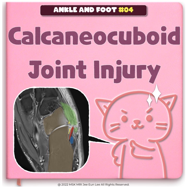
https://www.instagram.com/msk_mri/ The calcaneocuboid joint is stabilized by the dorsal calcaneocuboid ligament, short and long plantar ligaments, and calcaneocuboid component of the bifurcate ligament. ​​The calcaneocuboid joint capsule is reinforced dorsolaterally by the dorsal calcaneocuboid ligament The long and short plantar ligaments (sometimes referred to together as the plantar or inferi..

Ankle MRI 2022.12.18

[Ankle and Foot_02] Two fascicles of the anterior talofibular ligament (ATFL)

The anterior talofibular ligament (ATFL) is composed of the superior (S-ATFL) and inferior fascicle (I-ATFL). Both fascicles have a contiguous footprint on the anterior distal fibula border, however, their distal insertion on the talus is located apart from each other. The superior fascicle of the ATFL is a distinct anatomical structure, whereas the inferior ATFL fascicle and the CFL share some ..

Ankle MRI 2022.12.12

[Ankle and Foot_01] anterior talofibular ligament (ATFL), and posterior talofibular ligament (PTFL)

The lateral ligament complex of the ankle consists of the anterior talofibular ligament (ATFL), calcaneofibular ligament (CFL), and posterior talofibular ligament. This ligament plays an important role in limiting anterior displacement, internal rotation of the talus, and plantar flexion of the ankle The anterior talofibular ligament extends from the anterolateral malleolar tip to the talar neck..

Ankle MRI 2022.12.11

[Ankle/foot_Case_#05] Fracture through the ATFL talar tunnel, lateral ankle ligament reconstruction

When instability persists despite an anatomic repair, many surgeons turn to anatomic lateral ankle ligament reconstruction with autogenous or allogenic tendons. Several techniques have been described and most require the creation of two transosseous tunnels in the distal fibula, one for the ATFL and one for the CFL. The popularity of these procedures has been increasing over the last decade with..

Ankle MRI/[Case] 2022.09.18

[Ankle/foot_Case_#05] [Easy level] Normal anatomy and ATFL complete tear, CFL partial tear

ATFL The ATFL is best imaged on axial T1 or high-resolution PD MR images, appearing as a flat, thin, homogeneous band of low signal intensity arising from the anterior margin of the lateral malleolus and coursing anteromedially downward to attach onto the neck of the talus, just anterior to the fibular articular cartilage. The CFL is large, strong, and cordlike. It arises from the deep aspect of..

Ankle MRI/[Case] 2022.09.18

[Ankle/foot_Case_#04] [Moderate level] Injury of flexor retinaculum, Fascial Sleeve of the Medial Malleolus

The flexor retinaculum is often torn in conjunction with a tear of the superficial deltoid ligament, or in association with ankle fracture. Tears of the flexor retinaculum have been described in association with tibialis posterior tendon dislocation. An isolated tear of the flexor retinaculum is rare. “Fascial sleeve of the medial malleolus,” is composed of the superficial deltoid origin, the me..

Ankle MRI/[Case] 2022.09.16

[Ankle/foot_Case_#03] [Easy level] Achilles tendon complete rupture, Typical case

The spectrum of tears ranges from microtears to interstitial tears (parallel to the long axis of the Achilles), to partial tears, and eventually to complete tears (ruptures). Typically, in a young 'normal' individual, the Achilles tendon ruptures in the 'critical zone’, which is a region of relative watershed hypovascularity 2-6 cm proximal to insertion. The Achilles tendon tear classification i..

Ankle MRI/[Case] 2022.09.16

[Ankle/foot_Case_#02] [Challenging level] Mueller-Weiss syndrome

#AnkleMRI, #Mueller-Weiss, #Hindfootvarus, #Navicular, #footpain, #footosteoarthritis, #chroicfootpain #안산에이스병원,#Ansan_Ace_Hospital, #MSK_MRI, #mskmri_Ankle, #영상의학과이지은, #영상의학공부맛집, Mueller Weiss syndrome is a spontaneous adult onset osteonecrosis of the tarsal navicular. This syndrome is multifactorial and related to chronic loading on a suboptimally ossified navicular bone susceptible to central..

Ankle MRI/[Case] 2022.09.11

[Ankle_01_Instagram] Medial Plantar Neuropathy, Jogger’s foot, Knot of Henry, fo

Jogger’s foot can develop at the Henry knot owing to repetitive trauma to the nerve, especially while running with increased heel valgus and foot pronation. Patients usually present with heel and arch pain, tenderness and a positive Tinel sign posterior to the navicular tuberosity, and numbness along the medial plantar foot. • MR: Edema or atrophy of the abductor hallucis and flexor digitorum br..

Ankle MRI 2022.05.29

[Ankle_01_Review] Medial Plantar Neuropathy, Jogger’s foot, Knot of Henry, foot

카페 회원이 되시면 pdf로 보실 수 있습니다. MSK MRI와 즐겁게 공부하고 공유하는 영상의학과 의사들의 모임 https://cafe.naver.com/studywithmskmri/ 근골격 영상의학에 관심이 많은 영상의학 선생님들과 함께 공부하고 성장하는 채널입니다. 영상의학 선생님들(전문의, 전공의) 모두 환영합니다. 함께 즐겁게 공부하는 공간이 되었으면 합니다. 여기 동영상들은 모두 비상업적이며, 오직 교육 목적으로 만든 것임을 분명히 밝힙니다. 그러나 이 동영상의 무단편집 및 재가공, 혹은 영상의 공유는 절대금지합니다. #MSKMRI, #virtualMRI, #radiologist, #musculoskeletal #AnkleMRI, #FootMRI, #MSKMRI_ANKLE #Denervatio..

Ankle MRI 2022.05.29