본문 바로가기

meniscaltear4

(Fig 1-B.02) Horizontal Meniscal Tears 
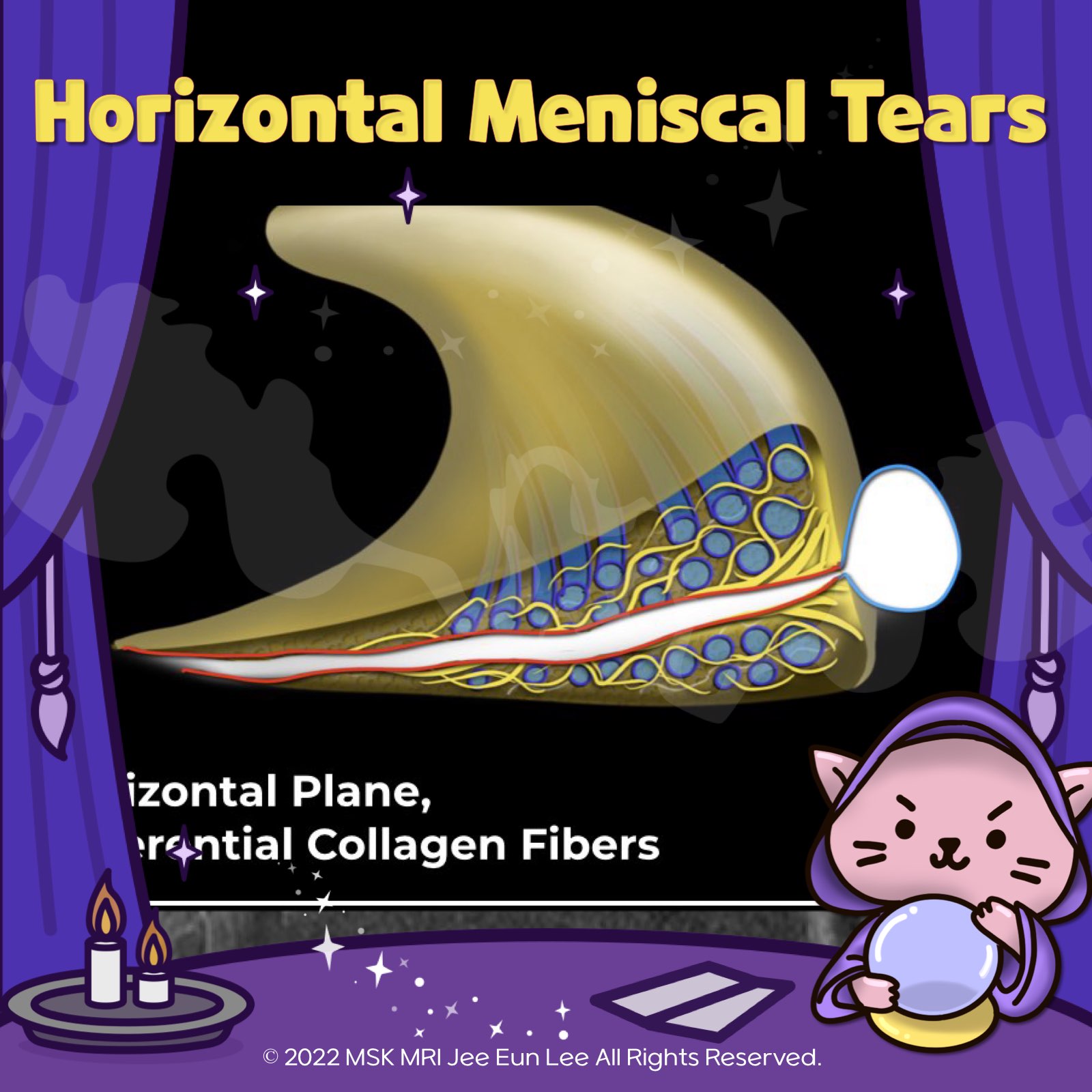
https://youtu.be/CD8P75631LI https://youtu.be/nKQsItyKAtM Horizontal Tear Meniscal tear in horizontal plane, dissecting through circumferential collagen fibers. MRI Appearance of Tear Appears as a horizontally oriented line of increased intrameniscal signal extending to the superior or inferior surface of the meniscus, typically near the free edge. Common Location Most common within the posterio.. 2024. 1. 17.
(Fig 1-A.17) Oblique meniscomeniscal ligament https://youtu.be/fyX1V1RrctQ https://youtu.be/kEuMzUrHD0M Aspect Details Ligament Type Oblique Meniscomeniscal Ligaments (Intermeniscal Ligament) Prevalence 1% to 4% Location Transverse the intercondylar notch between the anterior and posterior cruciate ligaments Naming Based on anterior attachment sites Medial OMML Originates from the anterior horn of the medial meniscus extends through the int.. 2024. 1. 15.
(Fig 1-A.06) Medial Meniscus Posterior Root, Anatomy, degeneration, tear https://youtu.be/CjYENTgOT3Y https://youtu.be/kEuMzUrHD0M "🧐 Deep Dive into the Meniscofemoral Ligament (MFL) Anatomy! ✨ The MFL, a critical component of the knee, originates from the posterior horn of the lateral meniscus and inserts onto the lateral aspect of the posterior medial femoral condyle. It plays a key role in the stability and function of the knee joint. 🔍 Let's break it down: 1️⃣ Th.. 2024. 1. 7.
(Fig 1-A.06) Medial Meniscus Posterior Root, Anatomy, degeneration, tear https://youtu.be/GvR9ULGrDOw https://youtu.be/kEuMzUrHD0M Category Description Insertion Points - Inserts along the downward slope of the intercondylar fossa, anterior to the posterior cruciate ligament. - Extends from the tibial attachment point to just lateral from the articular cartilage inflection point of the medial tibial plateau. - Commences just lateral to the apex of the medial tibial p.. 2024. 1. 5.