본문 바로가기

mri anatomy14

[Anatomy_31] Anterior meniscofemoral ligament, ​​Anomalous insertion of the medi 
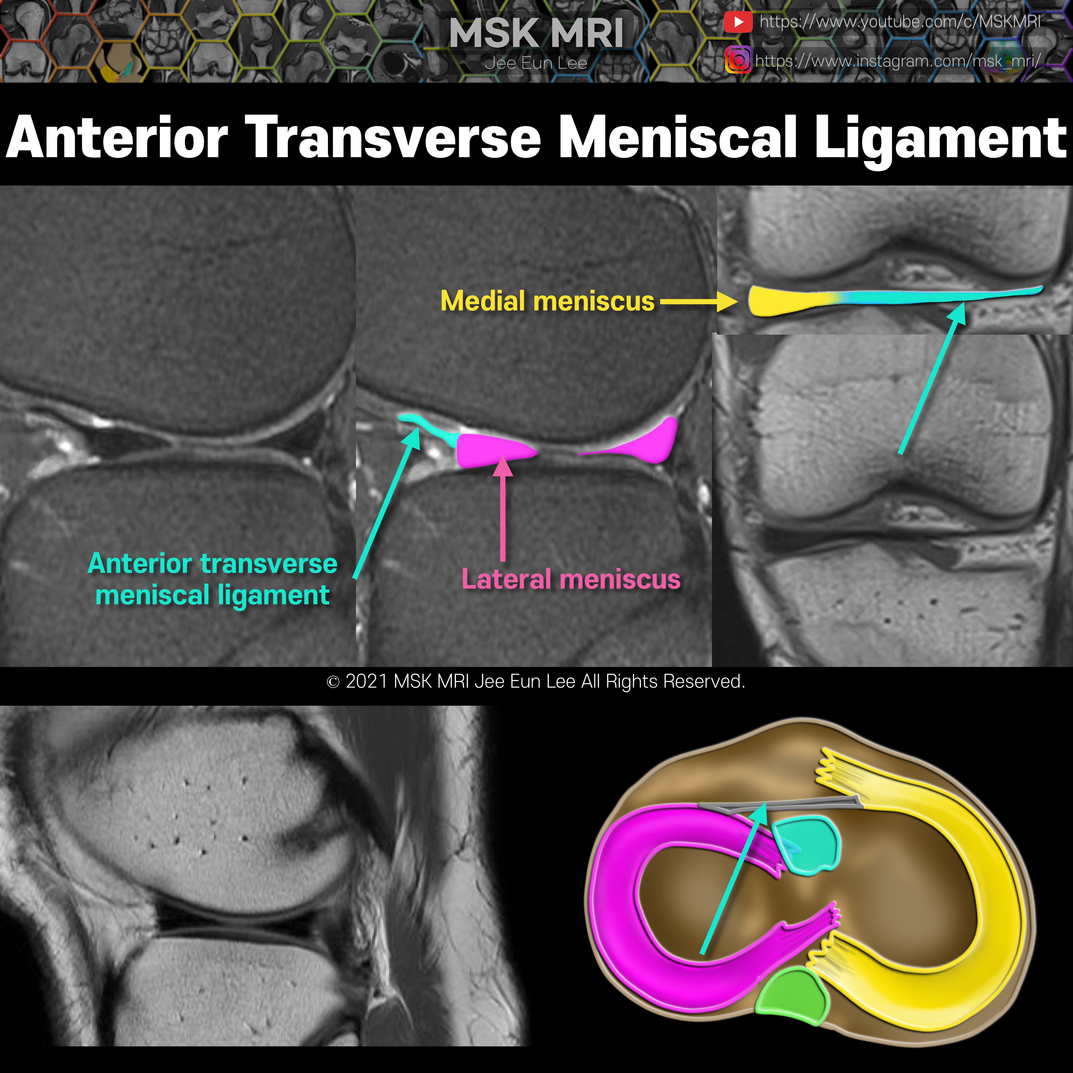
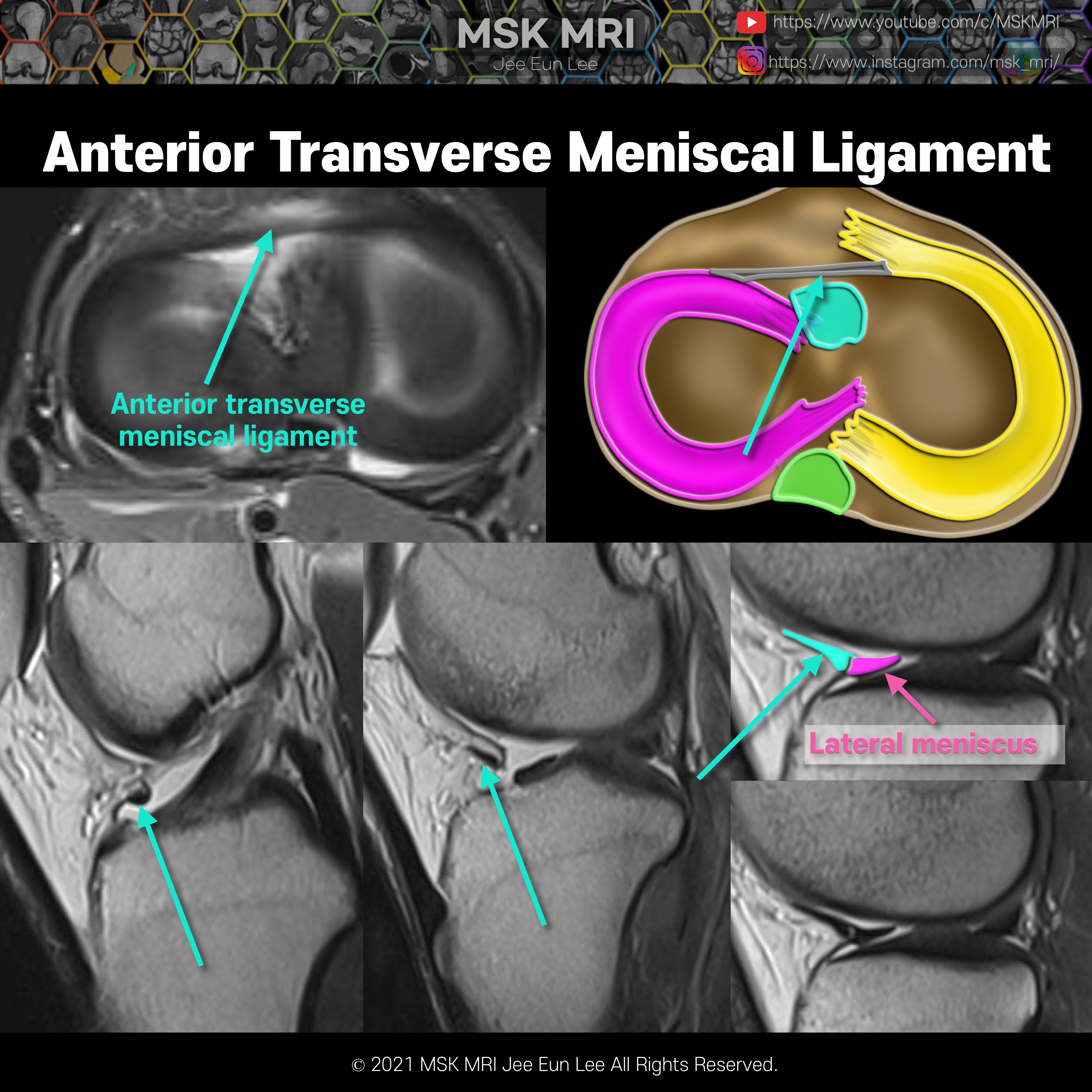
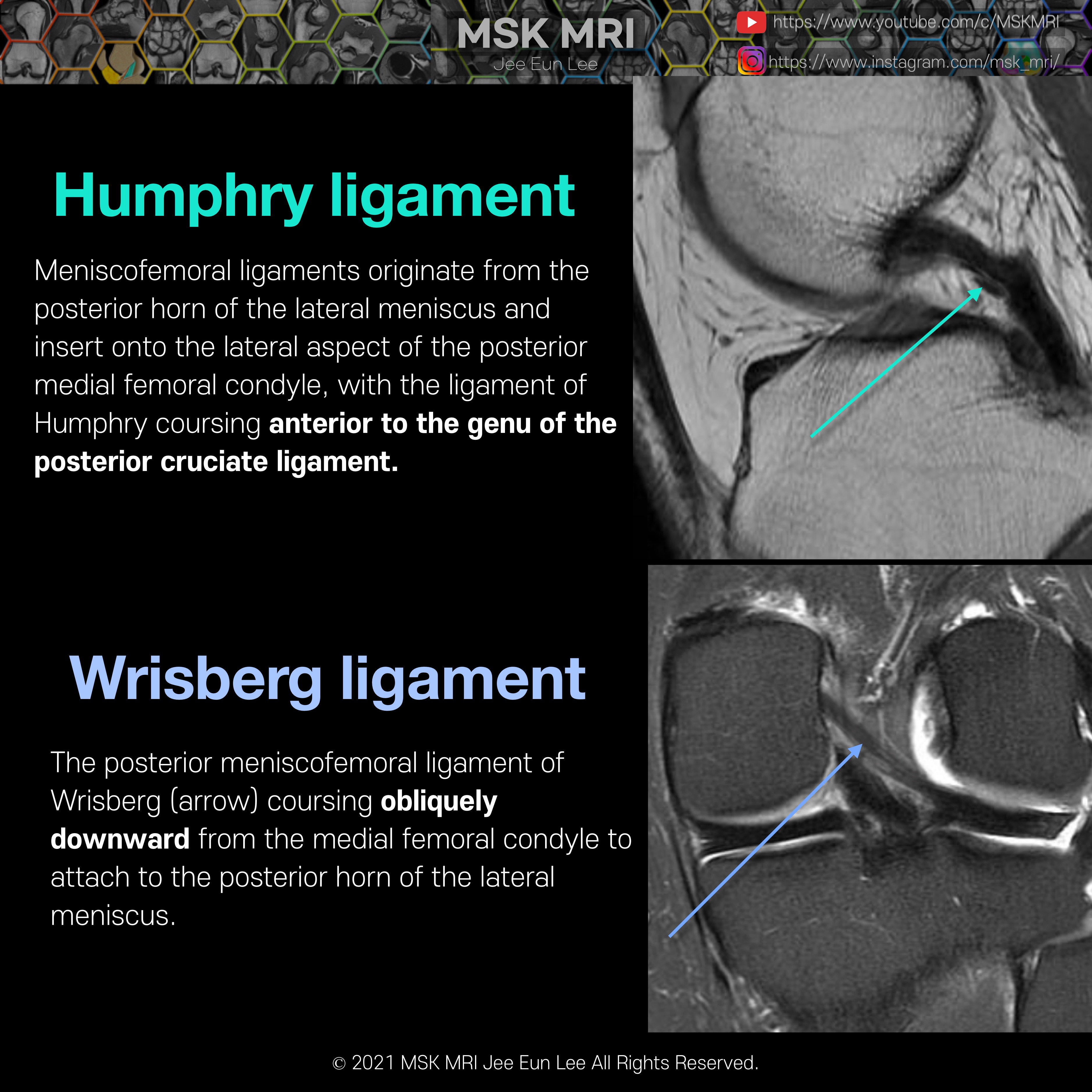
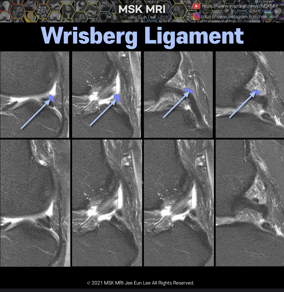
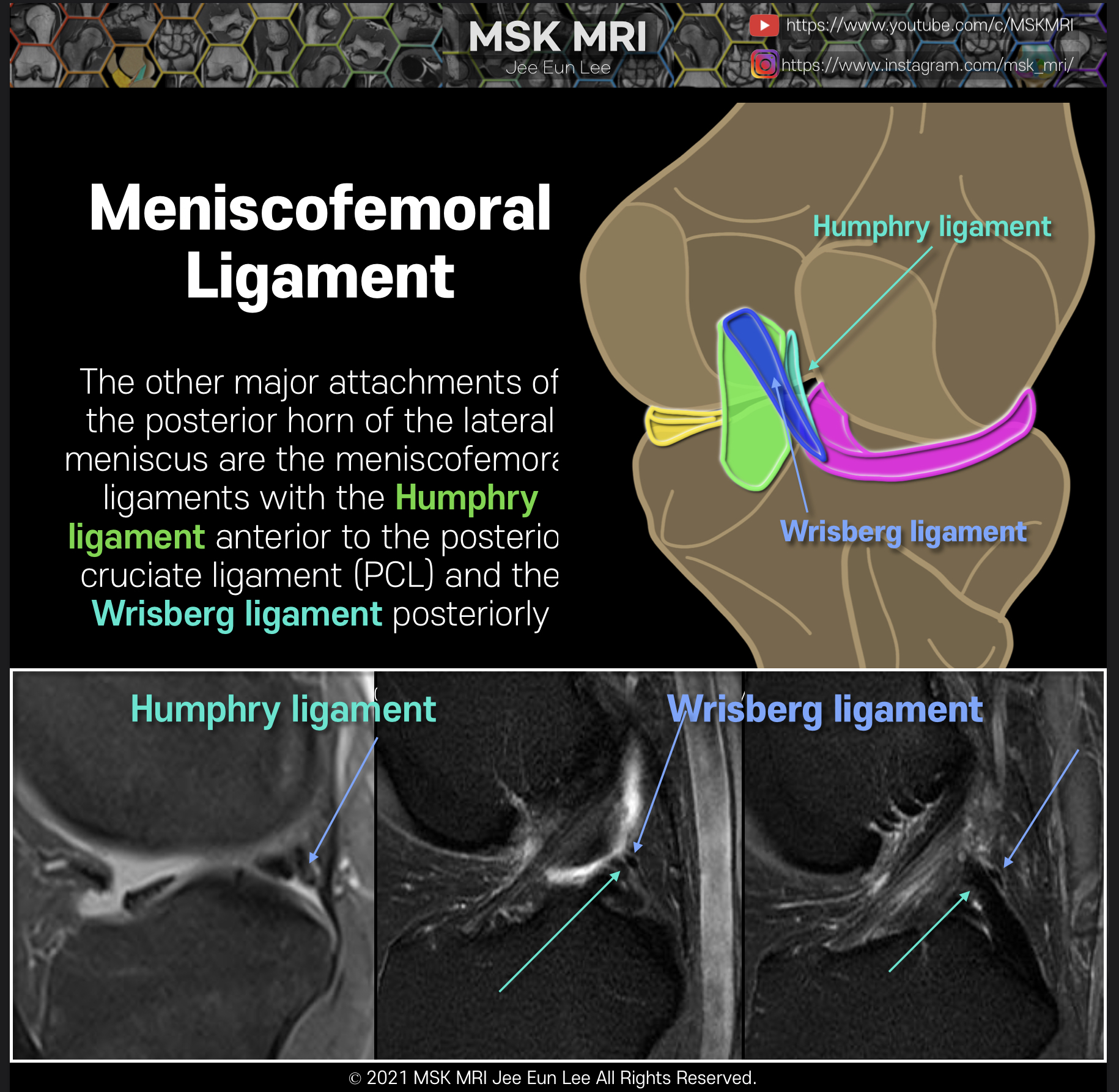
Anomalous insertion of the medial meniscus (AIMM) into the anterior cruciate ligament (ACL) is rare.The AIMM has the same anatomical description as the anteromedial meniscofemoral ligament It appears as a linear band isointense to the ACL and meniscus, which runs parallel to the ACL and demonstrates a hyperintense gap between it and the ACL On MRI, this anomaly may be misinterpreted as an ACL te.. 2021. 10. 2.
[Anatomy_30] Oblique Meniscomeniscal Ligament vs bucket handle tear -03 The medial OMML originates from the anterior horn of the medial meniscus and inserts onto the posterior horn of the lateral meniscus between the ACL and PCL. This structure should not be mistaken for a bucket handle meniscal tear A bucket-handle tear is a longitudinal tear with central migration of the inner “handle” fragment The correct diagnosis of an oblique meniscomeniscal ligament rather th.. 2021. 10. 2.
[Anatomy_29] Oblique Meniscomeniscal Ligament -02 The medial OMML originates from the anterior horn of the medial meniscus and extends through the intercondylar notch to insert onto the posterior horn of the lateral meniscus. This ligament passes between the ACL and PCL. This oblique meniscomeniscal ligament is kind of similar SI to meniscal tissue. The sagittal image near the midline demonstrates a linear low signal intensity structure atop th.. 2021. 10. 2.
[Anatomy_28] Oblique Meniscomeniscal Ligament -01 The medial and lateral oblique menisco-meniscal ligaments are rare and can mimic a tear if not recognized. The oblique meniscomeniscal ligament passes from the anterior horn of one meniscus to the posterior horn of the opposite meniscus. The oblique meniscomeniscal ligaments are named based on their anterior attachment site. The medial OMML originates from the anterior horn of the medial meniscu.. 2021. 10. 2.
[Anatomy_27] Anterior transverse meniscal ligament -02 Both horns of the lateral meniscus are triangular in shape and have very sharp points. In this case, we can this low-signal-intensity band like structure adjacent to the anterior horn of the lateral meniscus Both cases are normal finding, The transverse ligament has various diameters. The anterior root of MM inserts along the tibial eminence, and posterior fibers of the anterior horn attachment .. 2021. 9. 27.
[Anatomy_26] Anterior transverse meniscal ligament -01 The transverse ligament is a fibrous band that connects the anterior horns of the medial and lateral menisci. Axial and sagittal images demonstrate the course of the transverse ligament as a low-signal-intensity band traversing Hoffa’s infrapatellar fat pad. The transverse ligament can simulate an oblique tear adjacent to the anterior horn of the lateral meniscus © 2021 MSK MRI Jee Eun Lee All R.. 2021. 9. 27.
[Anatomy_24] Meniscofemoral ligament, Humphry ligament The meniscofemoral ligaments originate from the posterior horn of the lateral meniscus and insert onto the lateral aspect of the posterior medial femoral condyle, anterior to the PCL. notice, This cleft between the meniscus and meniscofemoral ligament may be mistaken for a meniscal tear. © 2021 MSK MRI Jee Eun Lee All Rights Reserved. #MSKMRI, #virtualMRI, #radiologist, #Knee_MRI, #MSKMRI_Knee, .. 2021. 9. 26.
[Anatomy_23] Meniscofemoral ligament, Wrisberg ligament, PseudoTear, Wrisberg Ri https://youtu.be/twVB-GMvcIg Coronal image demonstrates the posterior meniscofemoral ligament of Wrisberg (arrow) coursing obliquely downward from the medial femoral condyle to attach to the posterior horn of the lateral meniscus The meniscofemoral ligaments originate from the posterior horn of the lateral meniscus and insert onto the lateral aspect of the posterior medial femoral condyle, poste.. 2021. 9. 26.
[Shorts #05] Meniscofemoral ligament, Wrisberg ligament #shorts •This posteroblique illustration shows the relationship of meniscofemoral ligaments, lateral meniscus, and PCL. The meniscofemoral ligaments originate from the posterior horn of the lateral meniscus and insert onto the lateral aspect of the posterior medial femoral condyle, with the Humphry ligament anterior to the posterior cruciate ligament (PCL) and the Wrisberg ligament posteriorly Subset of.. 2021. 9. 26.
[Anatomy_22] Meniscofemoral ligament, Humphry, Wrisberg The major attachments of the posterior horn of the lateral meniscus are the popliteomeniscal fascicles and the meniscofemoral ligaments. This posteroblique illustration shows the relationship of meniscofemoral ligaments, lateral meniscus, and PCL. The meniscofemoral ligaments originate from the posterior horn of the lateral meniscus and insert onto the lateral aspect of the posterior medial femo.. 2021. 9. 26.
[Shorts #03] Meniscofemoral ligament, Humphry, Wrisberg #shorts •This figure illustrates the relationship of meniscofemoral ligaments, lateral meniscus, and PCL. The meniscofemoral ligaments originate from the posterior horn of the lateral meniscus and insert onto the lateral aspect of the posterior medial femoral condyle, with the Humphry ligament anterior to the posterior cruciate ligament (PCL) and the Wrisberg ligament posteriorly Occasionally the menisc.. 2021. 9. 25.
[Anatomy_21] Posteroinferior popliteomeniscal fascicle The popliteomeniscal fascicles consist of the anteroinferior(strongest), posterosuperior, and posteroinferior. posteroinferior The posteroinferior popliteomeniscal fascicle (arrow) is the least consistently visualized of the fascicles The posteroinferior fascicle is more medially located, and extends from a medial aponeurotic extension of the popliteus tendon to attach to the lateral meniscus ne.. 2021. 9. 25.
[Anatomy_20] Posterosuperior popliteomeniscal fascicle The popliteomeniscal fascicles arise from the periphery of the posterior horn of the lateral meniscus and form a posterolateral meniscocapsular extension, which creates the popliteus hiatus. The popliteal hiatus shows relatively symmetric posterosuperior and anteroinferior fascicles. More medial section of the hiatus shows the normal deficiency of the anteroinferior popliteomeniscal fascicle. le.. 2021. 9. 25.
[Anatomy_18] Anteroinferior popliteomeniscal fascicle 01 The anteroinferior popliteomeniscal fascicle (yellow arrow) is seen to course from the inferior aspect of the lateral meniscus posteroinferiorly to blend with fibers of the popliteofibular ligament This fascicle is typically seen on sagittal slices where the fibular head is also visible. In another patient, a component of the anteroinferior popliteomeniscal fascicle which courses directly to the.. 2021. 9. 25.