pmf2 (Fig 1-A.13) normal popliteomeniscal fascicle deficiency 
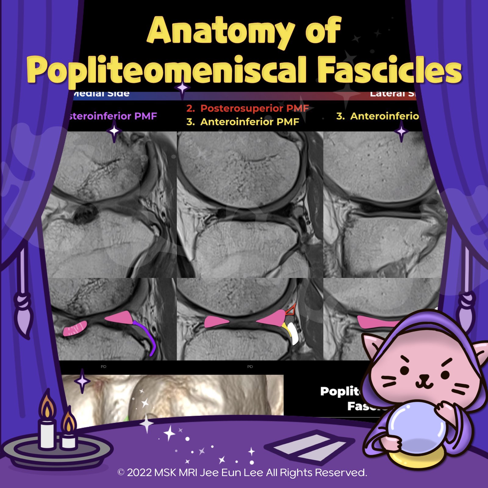
https://youtu.be/ckDHFabBkUk?si=01vgw2dU-6veGl1S https://youtu.be/kEuMzUrHD0M 👉Anteroinferior Popliteomeniscal Fascicle: This fascicle forms the lateral floor of the popliteal hiatus, varying in thickness. It connects the lateral meniscus’s body, extends to the popliteus muscle’s musculotendinous unit, and blends with the popliteofibular ligament, inserting at the fibular head’s styloid process... 2024. 1. 11. (Fig 1-A.12) Anatomy of popliteomeniscal fascicles https://youtu.be/GYwJcbKhxbY Fascicle Location and Appearance Anteroinferior Popliteomeniscal Fascicle Visible on sagittal slices near the fibular head; extends posteroinferiorly from the lateral side of the lateral meniscus (LM) and merges with the popliteus tendon; conjoined attachment with the popliteofibular ligament at the fibula’s styloid process; variable appearance. Posterosuperior Popli.. 2024. 1. 10. 이전 1 다음