==============================================
⬇️✨⬇️🎉⬇️🔥⬇️📚⬇️
Click the link to purchase on Amazon 🎉📚
==============================================
🎥 Check Out All Videos at Once! 📺
👉 Visit Visualizing MSK Blog to explore a wide range of videos! 🩻
https://visualizingmsk.blogspot.com/?view=magazine
📚 You can also find them on MSK MRI Blog and Naver Blog! 📖
https://www.instagram.com/msk_mri/
Click now to stay updated with the latest content! 🔍✨
==============================================
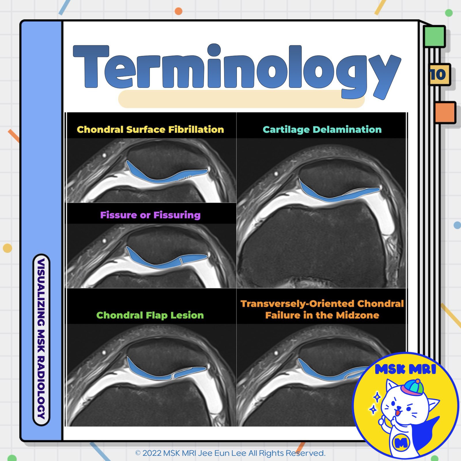
📌 Cartilage Damage Terminology
1️⃣. Fibrillation
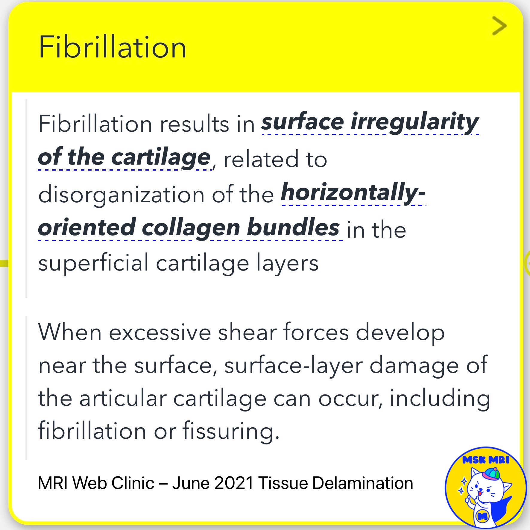
- Fibrillation results in cartilage surface irregularity, related to the disorganization of the horizontally oriented collagen bundles in the superficial cartilage layers.
- When excessive shear forces develop near the surface, surface-layer damage of the articular cartilage can occur, including fibrillation or fissuring.
2️⃣. Fissure or Fissuring
- A fissuring is defined as a long and narrow split or crack.
- Chondral fissuring occurs primarily as a result of delamination between the obliquely and vertically oriented collagen fibrils rather than by breakage of collagen fibrils.
- Chondral fissures that extend deep can propagate perpendicular to the bone.
- These lesions can be associated with reactive bone marrow edema that calls attention to the lesion and aids in detection with MRI.
3️⃣. Cartilage Delamination
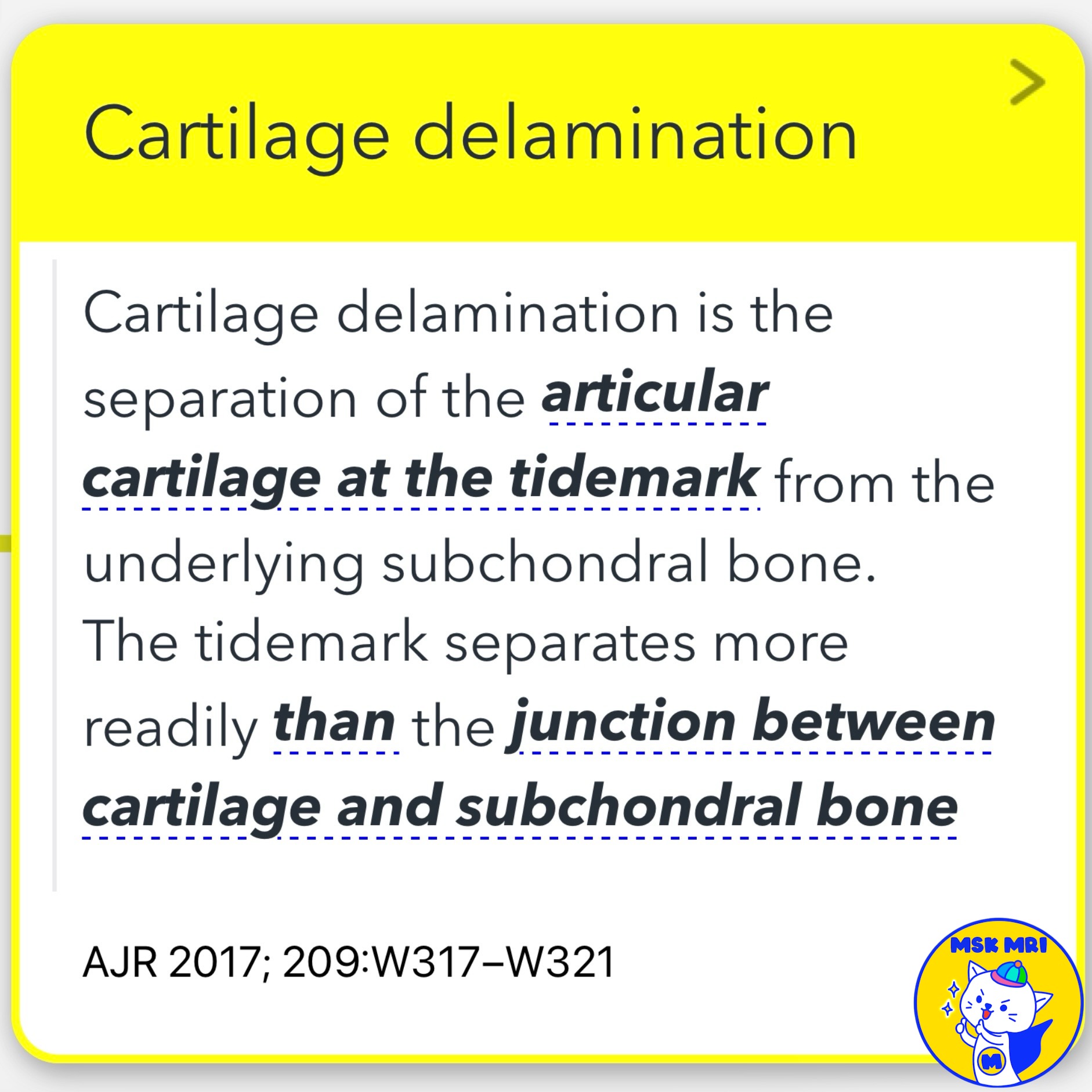
- Cartilage delamination separates the articular cartilage at the tidemark from the underlying subchondral bone.
- The tidemark separates more readily than the junction between cartilage and subchondral bone.
4️⃣. Chondral Flap Lesion
- Chondral fissures may result in delamination of the cartilage and chondral flap lesions if there is extension of the fissure parallel to the surface of the bone.
5️⃣. Transversely-Oriented Chondral Failure in the Midzone
- On MRI, a transversely oriented region of failure in the midzone of articular cartilage is sometimes seen in association with chondral fissures.
- The transversely oriented region of failure that is sometimes seen in the midzone of articular cartilage may represent delamination between pathologically disorganized collagen fibers.
- Unstable chondral flap delaminating portions of the superficial and transitional zone can also be observed.
References
- MRI Web Clinic – June 2021 Tissue Delamination
- J Knee Surg. 2020 Nov;33(11):1088-1099
- AJR 2017; 209:W317–W321
"Visualizing MSK Radiology: A Practical Guide to Radiology Mastery"
© 2022 MSK MRI Jee Eun Lee All Rights Reserved.
No unauthorized reproduction, redistribution, or use for AI training.
#CartilageDamage, #Fibrillation, #Fissuring, #CartilageDelamination, #ChondralFlapLesion, #MRI, #BoneMarrowEdema, #ArticularCartilage, #Orthopedics, #KneeSurgery
'✅ Knee MRI Mastery > Chap 5AB. Chondral and osteochondral' 카테고리의 다른 글
| (Fig 5-A.12) Fissure or Fissuring in Cartilage (0) | 2024.07.05 |
|---|---|
| (Fig 5-A.11) Hypointense Lesion in Cartilage (0) | 2024.07.05 |
| (Fig 5-A.09) Summary of MRI Findings in Cartilage Damage (1) | 2024.07.04 |
| (Fig 5-A.08) International Cartilage Repair Society grade 3 and 4 (0) | 2024.07.04 |
| (Fig 5-A.07) Partial Thickness Cartilage Damage (0) | 2024.07.04 |


