Click the link to purchase on Amazon 🎉📚
==============================================
🎥 Check Out All Videos at Once! 📺
👉 Visit Visualizing MSK Blog to explore a wide range of videos! 🩻
https://visualizingmsk.blogspot.com/?view=magazine
📚 You can also find them on MSK MRI Blog and Naver Blog! 📖
https://www.instagram.com/msk_mri/
Click to stay updated with the latest content! 🔍✨
==============================================
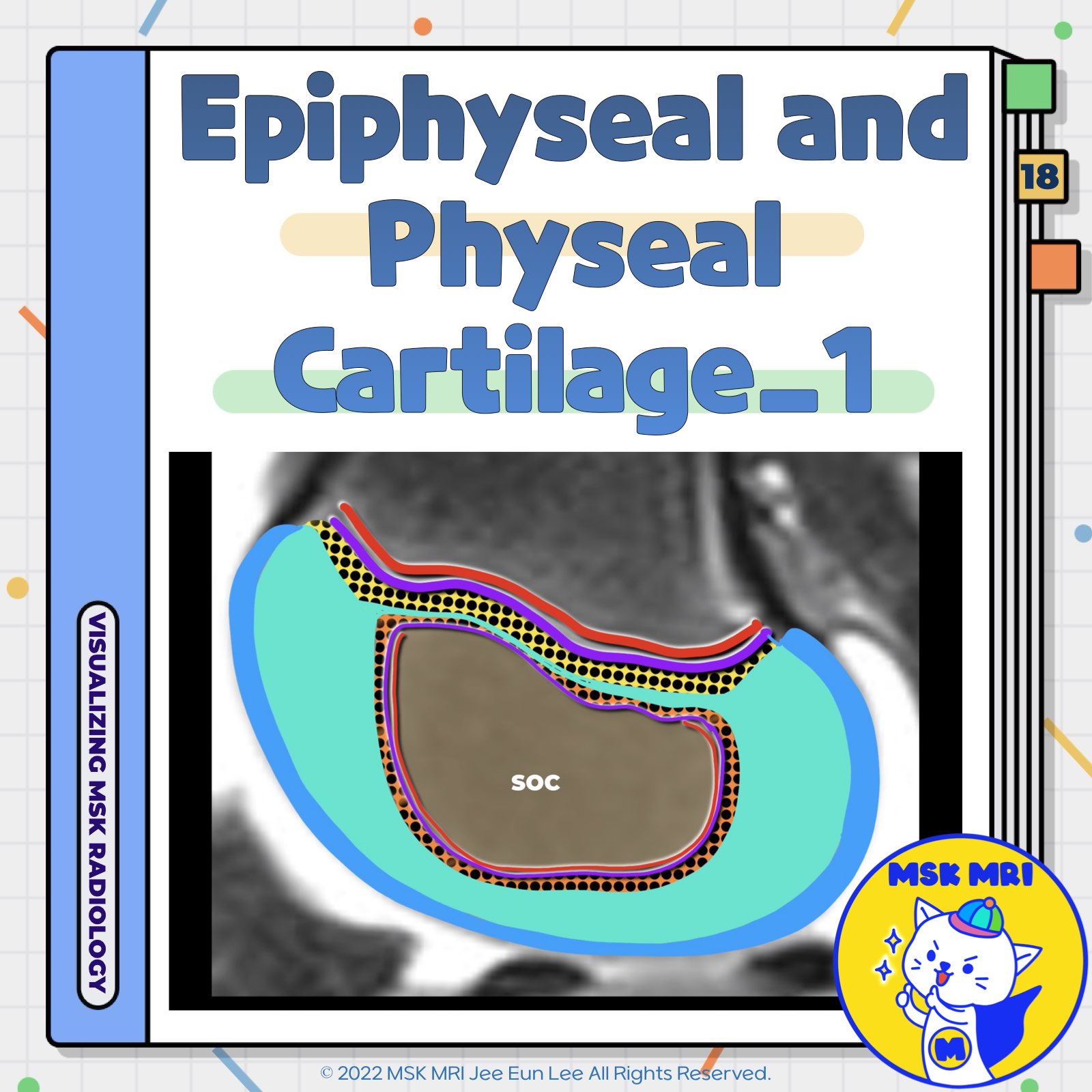
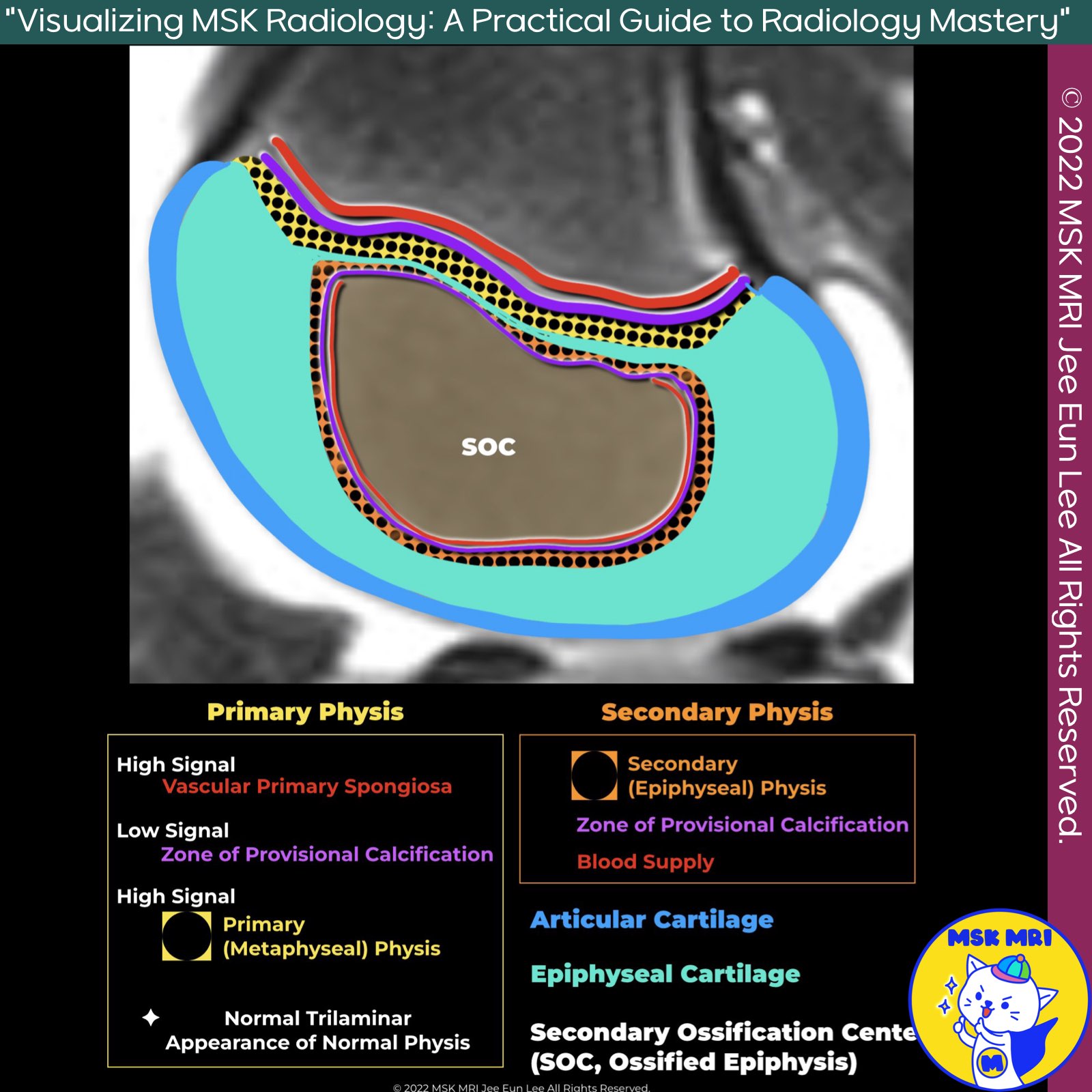
📌 Primary Physis and Secondary Ossification Center
✅ Introduction to Hyaline Cartilage
- Hyaline cartilage is highly cellular, constituting up to 75% of its volume.
- It plays a critical role in synthesizing new bone from cartilage, a process known as endochondral ossification.
✅ Primary Physis and Longitudinal Bone Growth
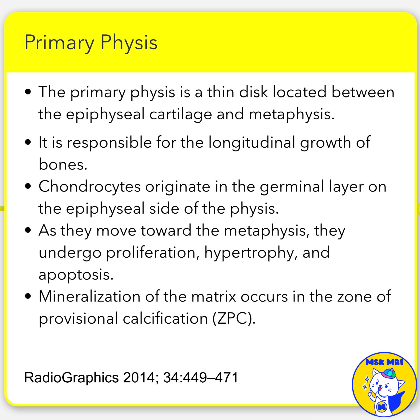
- The primary physis is a thin disk located between the epiphyseal cartilage and metaphysis.
- It is responsible for the longitudinal growth of bones.
- Chondrocytes in the physis are arranged in columns parallel to the long axis of the bone.
Chondrocyte Development and Ossification
- Chondrocytes originate in the germinal layer on the epiphyseal side of the physis.
- As they move toward the metaphysis, they undergo proliferation, hypertrophy, and apoptosis.
- Mineralization of the matrix occurs in the zone of provisional calcification (ZPC).
✅ Secondary Ossification Center (SOC)
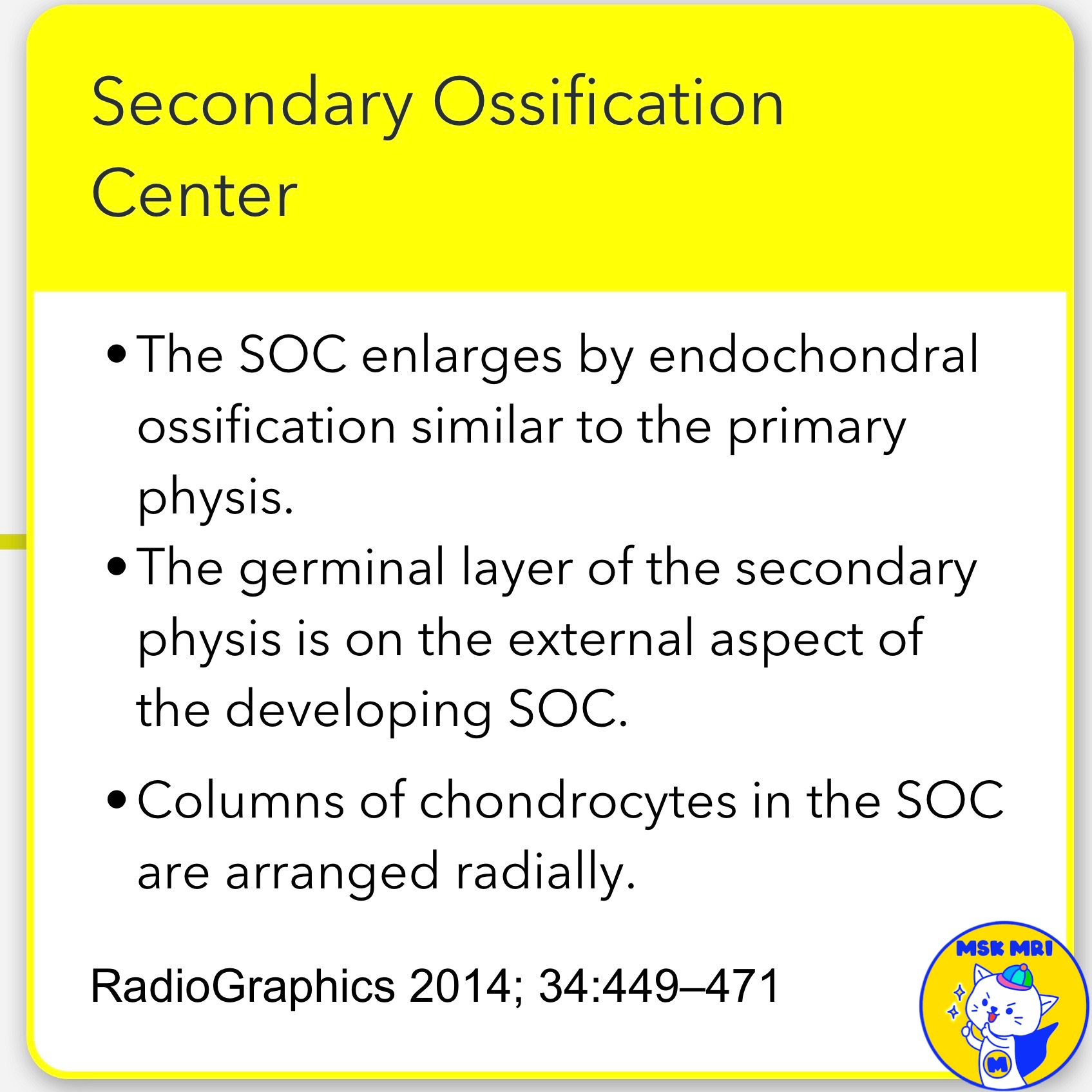
- The SOC enlarges by endochondral ossification similar to the primary physis.
- The germinal layer of the secondary physis is on the external aspect of the developing SOC.
- Columns of chondrocytes in the SOC are arranged radially.
References:
- RadioGraphics 2014; 34:449–471
"Visualizing MSK Radiology: A Practical Guide to Radiology Mastery"
© 2022 MSK MRI Jee Eun Lee All Rights Reserved.
No unauthorized reproduction, redistribution, or use for AI training.
#bonegrowth, #hyalinecartilage, #endochondralossification, #chondrocytes, #primaryphysis, #longitudinalgrowth, #provisionalcalcification, #secondaryossification, #SOC, #medicalresearch
'✅ Knee MRI Mastery > Chap 5AB. Chondral and osteochondral' 카테고리의 다른 글
| (Fig 5-B.20) Pathogenesis of Osteochondritis Dissecans (0) | 2024.07.13 |
|---|---|
| (Fig 5-B.19) Normal Epiphyseal and Physeal Cartilage: Part 2 (0) | 2024.07.13 |
| (Fig 5-B.17) Stress Response and Stress Fracture in Cancellous Bone (0) | 2024.07.11 |
| (Fig 5-B.16) Stress Response and Stress Fracture at Subchondral Level (0) | 2024.07.11 |
| (Fig 5-B.15) MRI Finding of Fracture Line (0) | 2024.07.11 |
