👉 Click the link below and request access—I’ll approve it for you shortly!
https://www.notion.so/MSKMRI-KNEE-b6cbb1e1bc4741b681ecf6a40159a531?pvs=4
==============================================
✨ Join the channel to enjoy the benefits! 🚀
https://www.youtube.com/channel/UC4bw7o0l2rhxn1GJZGDmT9w/join
==============================================
👉 "Click the link to purchase on Amazon 🎉📚"
[Visualizing MSK Radiology: A Practical Guide to Radiology Mastery]
https://www.amazon.com/dp/B0DJGMHMFS
==============================================
MSK MRI Jee Eun Lee
📚 Visualizing MSK Radiology: A Practical Guide to Radiology Mastery Now! 🌟 Available on Amazon, eBay, and Rain Collectibles! 💻 Ebook coming soon – stay tuned! ⏳ 🔗 https://www.amazon.com/dp/B0DJGMHMFS 🔗 https://www.ebay.com/itm/3875004193
www.youtube.com
Visualizing MSK Radiology: A Practical Guide to Radiology Mastery
www.amazon.com

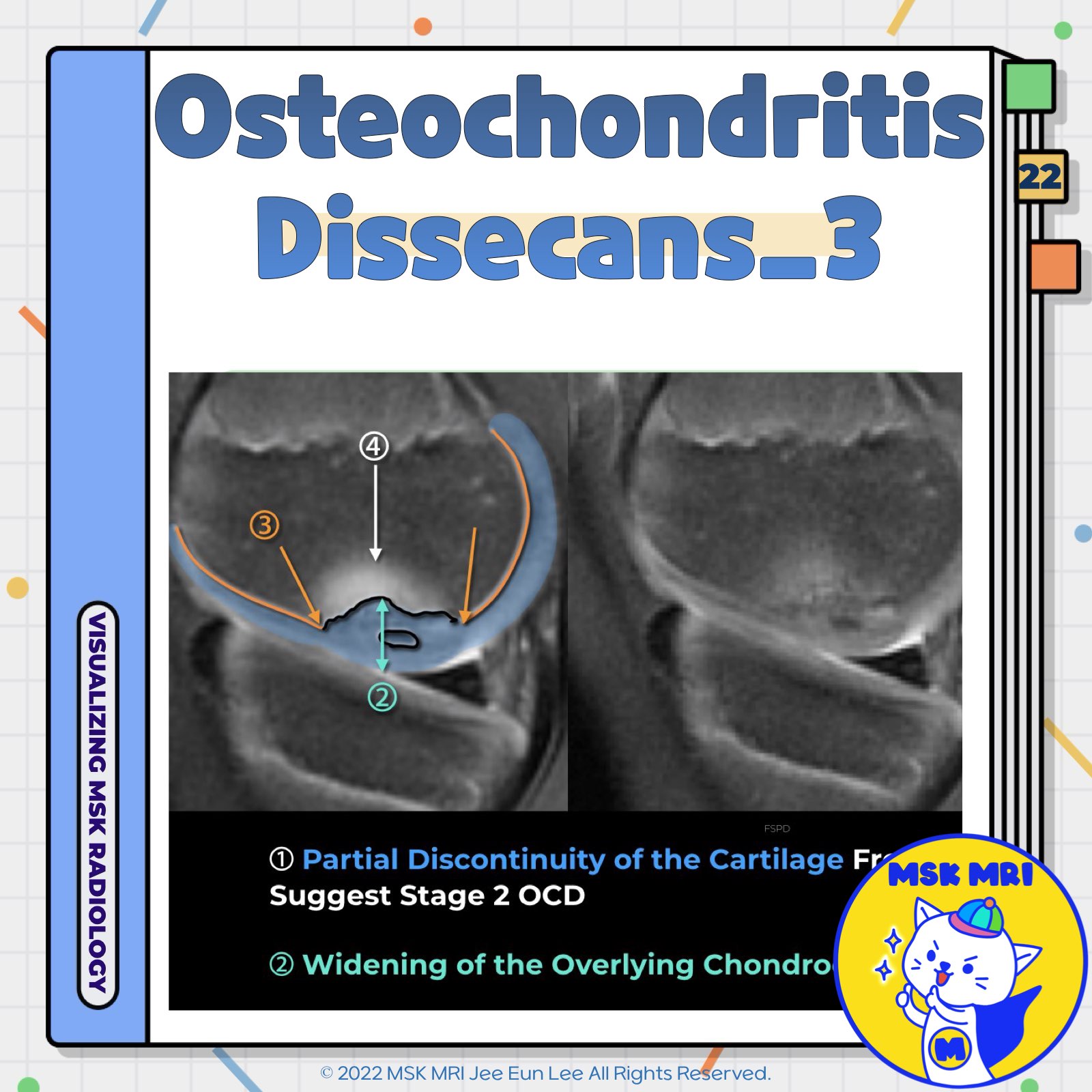
📌 MRI Findings of Osteochondritis Dissecans
✅ Key MRI Findings
- High Signal Intensity Rim
Observed at the interface between the fragment and the adjacent bone on T2-weighted sequences. - Fluid-filled Cysts
Presence of fluid-filled cysts beneath the lesion. - High Signal Intensity Line
A high signal intensity line extending through the articular cartilage overlying the lesion. - Focal Osteochondral Defect
A defect filled with joint fluid, indicating complete detachment of the fragment.
✅ Additional MRI Observations
- Subchondral Bone Irregularity
On MR images, OCD appears as an irregularity of the subchondral bone with underlying marrow edema-like signal intensity. - Secondary Physis Disruption in Children
MRI of children with OCD consistently showed secondary physis disruption, overlying chondroepiphyseal widening, and subchondral bone edema.
✅ Disease Progression
As the disease progresses, fragmentation and detachment of the subchondral bone are observed. Interestingly, this process of osteochondral fragmentation begins in the deep portion under the articular surface and eventually involves the superficial articular cartilage, suggesting an “inside-out” mechanism.
References
- Radiopaedia: Osteochondritis Dissecans
- Japanese Journal of Radiology (2022) 40:443–457
- AJR 2019; 213:963–982
- AJR 2012; 199:1121–1128
"Visualizing MSK Radiology: A Practical Guide to Radiology Mastery"
© 2022 MSK MRI Jee Eun Lee All Rights Reserved.
No unauthorized reproduction, redistribution, or use for AI training.
#OsteochondritisDissecans, #MRI, #Radiology, #SubchondralBone, #T2Weighted, #Cartilage, #BoneEdema, #PhysisDisruption, #PediatricRadiology, #MedicalImaging


'✅ Knee MRI Mastery > Chap 5AB. Chondral and osteochondral' 카테고리의 다른 글
| (Fig 5-B.24) Signs of Osteochondral Lesion Instability in Adults (0) | 2024.07.13 |
|---|---|
| (Fig 5-B.23) ICRS Staging System of Osteochondritis Dissecans (0) | 2024.07.13 |
| (Fig 5-B.21) Radiographic Findings of Osteochondritis Dissecans (1) | 2024.07.13 |
| (Fig 5-B.20) Pathogenesis of Osteochondritis Dissecans (0) | 2024.07.13 |
| (Fig 5-B.19) Normal Epiphyseal and Physeal Cartilage: Part 2 (0) | 2024.07.13 |