Click the link to purchase on Amazon 🎉📚
==============================================
🎥 Check Out All Videos at Once! 📺
👉 Visit Visualizing MSK Blog to explore a wide range of videos! 🩻
https://visualizingmsk.blogspot.com/?view=magazine
📚 You can also find them on MSK MRI Blog and Naver Blog! 📖
https://www.instagram.com/msk_mri/
Click to stay updated with the latest content! 🔍✨
==============================================
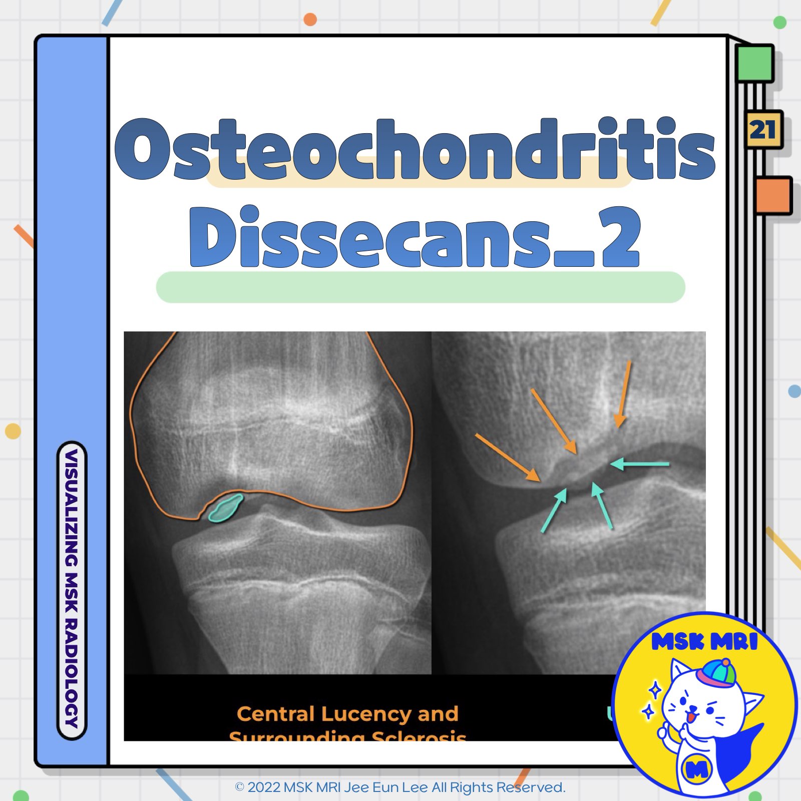
📌 Osteochondritis Dissecans
- Osteochondritis dissecans (OCD) is defined by the Research in Osteochondritis Dissecans of the Knee group as a "focal, idiopathic alteration of subchondral bone with risk of instability and disruption of adjacent articular cartilage that may result in premature osteoarthritis"
✅ Common Locations
- The classic and most common location of OCD in the knee is the lateral (intercondylar) aspect of the medial femoral condyle.
This area, along with the extended classic type (which also involves the central weight-bearing area), accounts for 75% of OCD cases.
- Lateral condylar lesions (13%)
- Medial central weight-bearing surfaces of the femoral condyle (10%)
- Patellar lesions
- Rarely, the anterior part of the femur (2%)
✅ Radiographic Findings
Early indications of OCD include subtle flattening or indistinct radiolucency above the cortical surface.
As the condition progresses, more pronounced symptoms such as:
- Contour abnormalities
- Fragmentation
- Density changes (both lucency and sclerosis)
In cases where an osteochondral fragment becomes unstable and displaced, both a donor site and intra-articular fragment may be observed.
References
- AJR 2019; 213:963–982
- RadioGraphics 2018; 38:1478–1495
- https://radiopaedia.org/articles/osteochondritis-dissecans
"Visualizing MSK Radiology: A Practical Guide to Radiology Mastery"
© 2022 MSK MRI Jee Eun Lee All Rights Reserved.
No unauthorized reproduction, redistribution, or use for AI training.
#OsteochondritisDissecans, #OCDKnee, #SubchondralBone, #ArticularCartilage, #PrematureOsteoarthritis, #MedialFemoralCondyle, #EarlyFindingsOCD, #RadiographicSigns, #OrthopedicResearch, #KneeHealth
'✅ Knee MRI Mastery > Chap 5AB. Chondral and osteochondral' 카테고리의 다른 글
| (Fig 5-B.23) ICRS Staging System of Osteochondritis Dissecans (0) | 2024.07.13 |
|---|---|
| (Fig 5-B.22) MRI Findings of Osteochondritis Dissecans (0) | 2024.07.13 |
| (Fig 5-B.20) Pathogenesis of Osteochondritis Dissecans (0) | 2024.07.13 |
| (Fig 5-B.19) Normal Epiphyseal and Physeal Cartilage: Part 2 (0) | 2024.07.13 |
| (Fig 5-B.18) Normal Epiphyseal and Physeal Cartilage: Part 1 (0) | 2024.07.13 |


